Bowen’s disease (squamous cell carcinoma of the skin in situ, squamous cell carcinoma of the skin in situ, CCM in situ) is a malignant neoplasm of the skin in the form of a hyperemic spot or plaque. Bowen’s disease is the very initial phase of squamous cell carcinoma of the skin when the cancerous growth of the basement membrane by malignant cells has not occurred. In this regard, it has a favorable prognosis and there is no likelihood of metastasis. Pathology occurs at the age of over 35-40 years, slightly more often in women.
Predisposing factors
There is no clear reason for the appearance of Bowen’s disease. It is only appropriate to talk about predisposing factors that, to varying degrees, can increase the risk of these neoplasms:
- Excessive insolation: excessive exposure to solar ultraviolet;
- Ionizing radiation;
- The influence of chemical compounds that damage the skin;
- Chronic skin injuries;
- Some dermatological diseases: Mibelli’s porokeratosis, dystrophic bullous epidermolysis, lichen planus, lupus erythematosus, Lewandowski-Lutz epidermodysplasia;
- The role of human papillomavirus in increasing the risk of Bowen’s disease is not ruled out.
Diagnostics
The diagnosis of Bowen’s disease is based on a clinical examination, which includes a routine examination of the formation and dermatoscopy. After examination, a biopsy is performed.
Symptoms
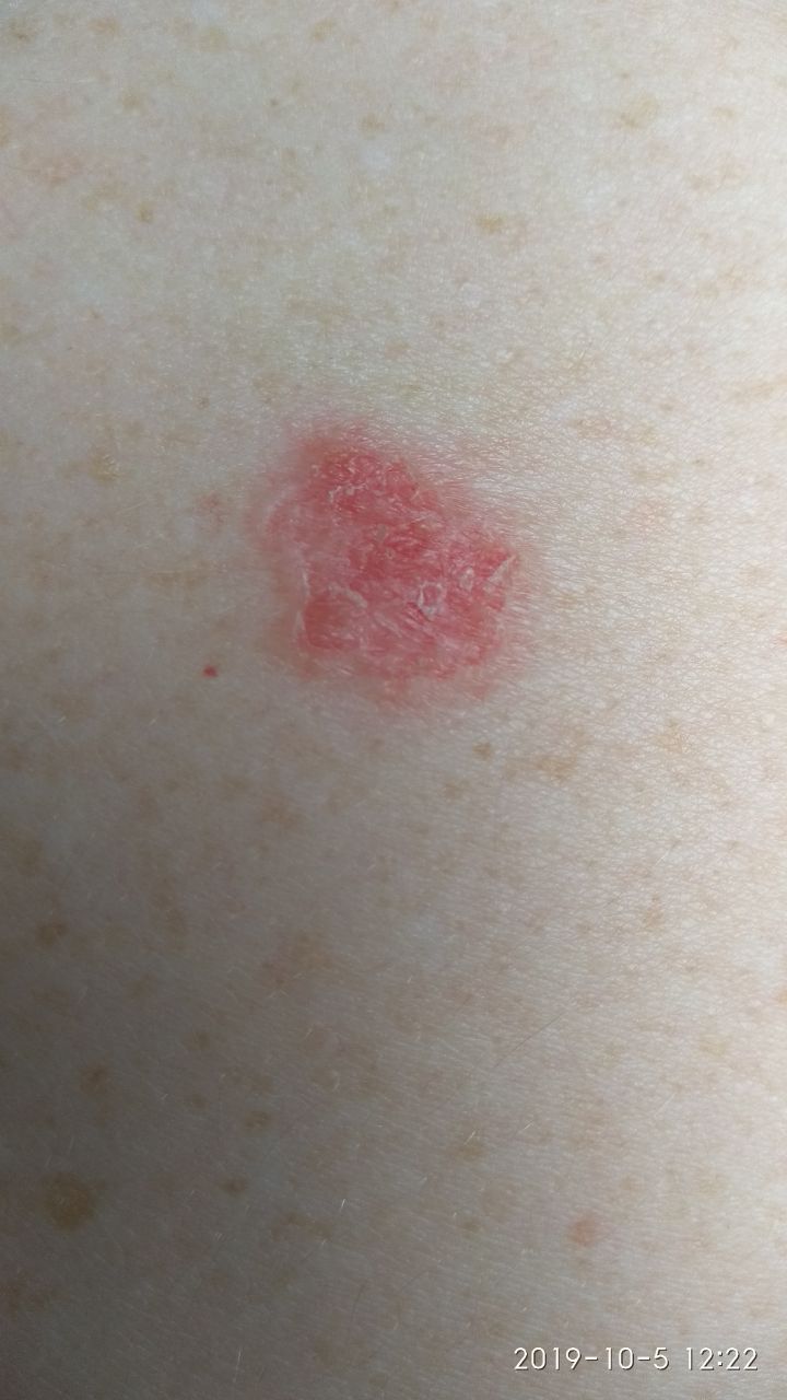
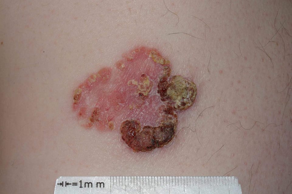
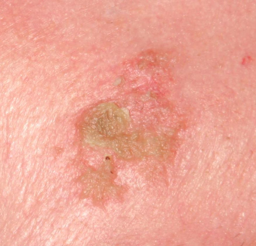
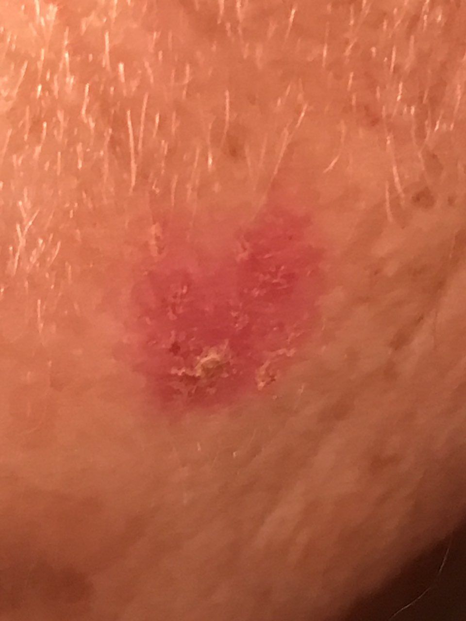
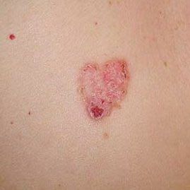
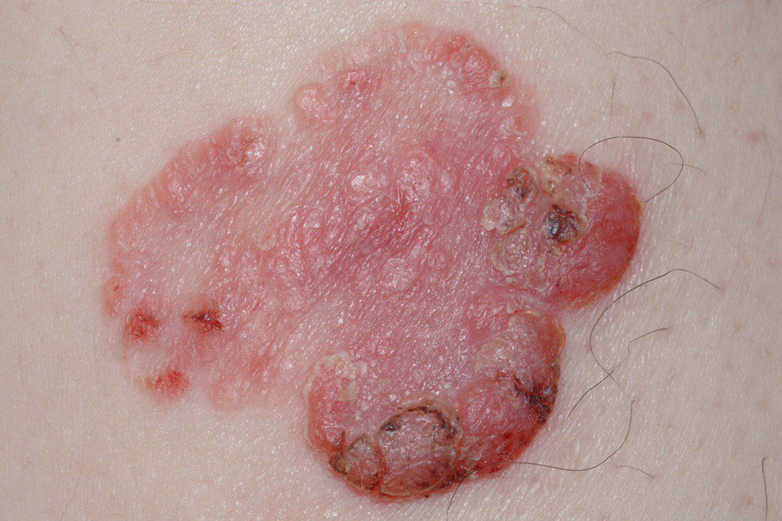
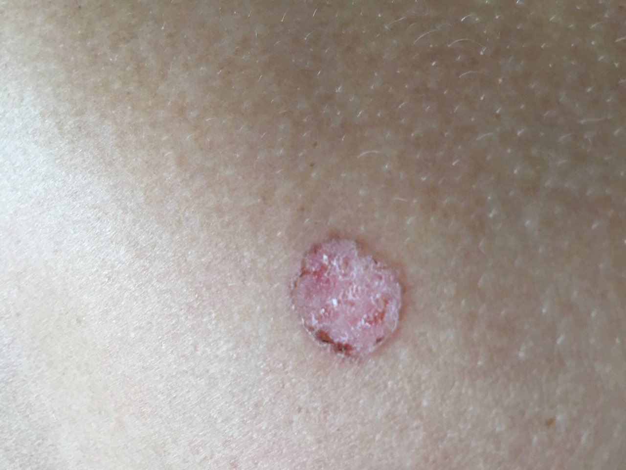
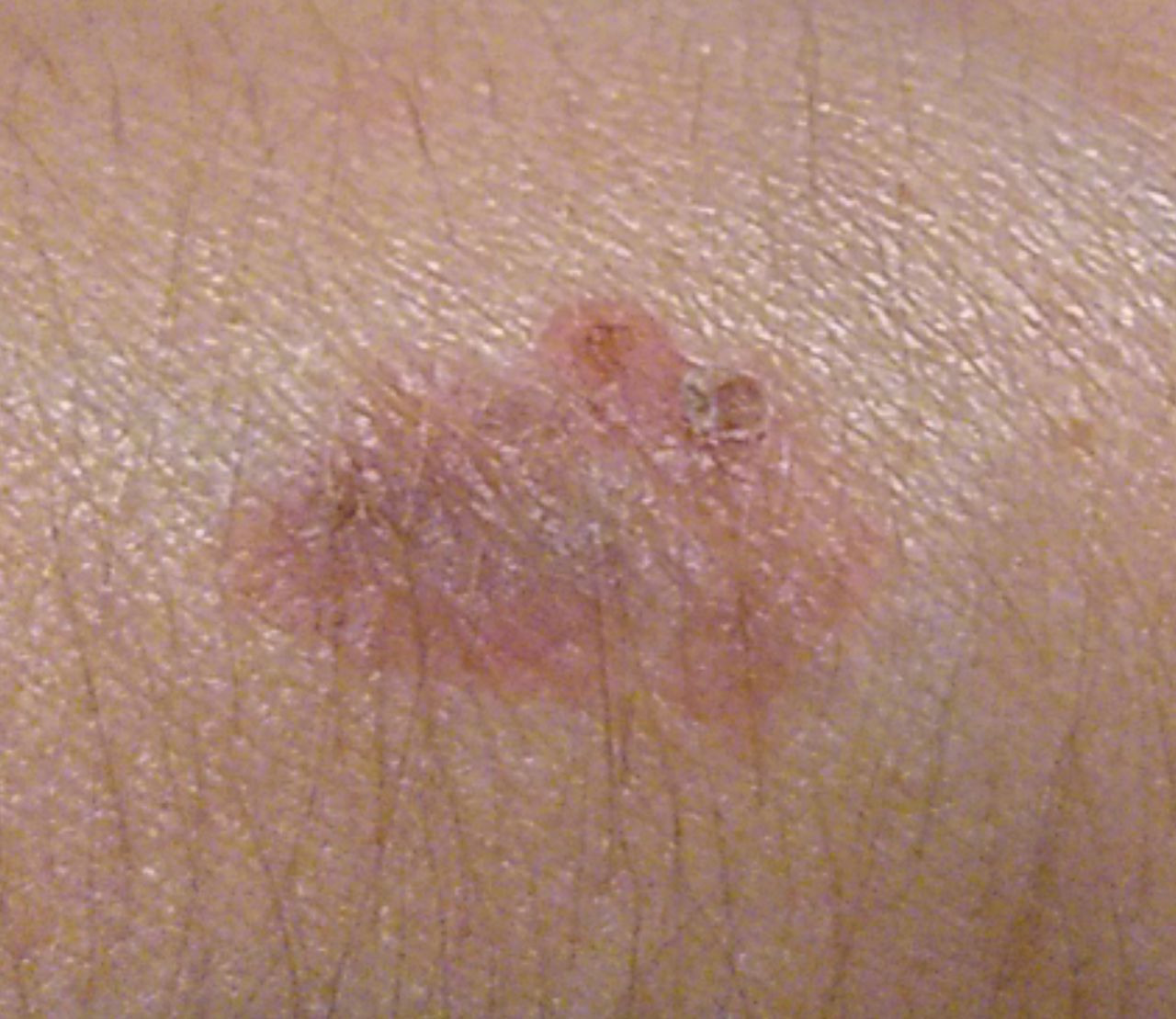
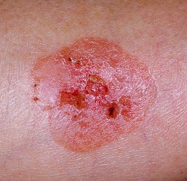
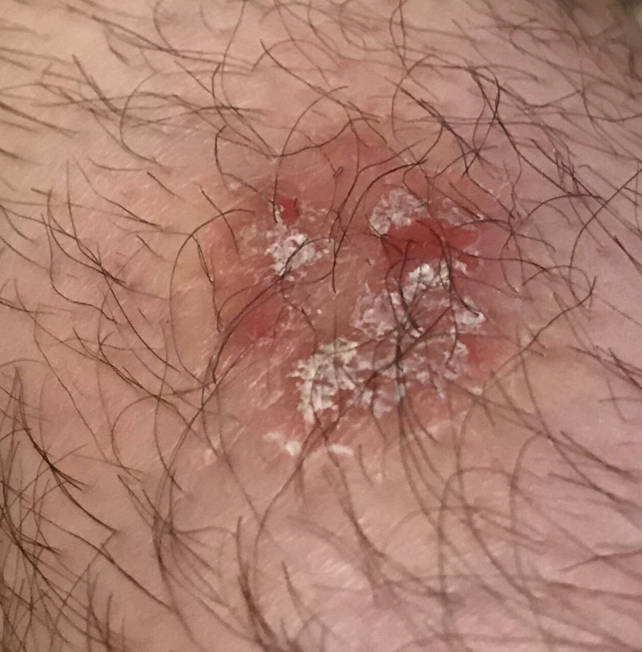
A visual examination of Bowen’s disease identifies single, rarely multiple, or grouped spots or plaques. The surface of the plaque differs significantly from the texture of ordinary skin: rough, tuberous, warty, focal covered with crusts, with erosion and signs of ulceration. It practically does not protrude above the skin (up to 1 mm, the edges of the tumor can protrude more).
The boundaries are usually clear, but uneven. The shape is irregular, asymmetric. The color is pink, pink-red, with the appearance of horny masses – gray shades join, the intensity of which depends on the severity of keratosis in the tumor area.
Hair growth is absent in the focal area.
Sizes range from 4 mm to 40 mm. Grouped structures from several foci can reach larger sizes. Increases slowly, grows over the years. Self-regression does not occur.
On palpation, in comparison with the surrounding skin, denser formation with peeling is determined. Crusts are usually easily removed, under them is eroded skin with hyperemia. Subjective sensations are usually absent or slight itching, burning.
The foci of Bowen’s disease are located on the torso. Also with great frequency, the face, scalp, neck, upper limbs (the area of the shoulder girdle, and hand) are affected.
Dermatoscopic Description
With dermatoscopy of foci of Bowen’s disease, the following features are visualized:
- A large number of vessels in the form of glomeruli;
- Point vessels on the background of a uniform pale red or pink color;
- Uniform distribution of vascular structures throughout the formation;
- Lack of pigmentation.
Differential diagnosis
Differential diagnosis is carried out with such pigmented neoplasms, as:
- Psoriasis, eczema, dermatitis;
- Seborrheic keratosis;
- Actinic keratosis;
- Lentigo melanoma;
- Basal cell carcinoma;
- Squamous cell carcinoma.
Risks
Bowen’s disease is a non-invasive squamous cell carcinoma of the skin (cancer in situ). The main difference from conventional skin carcinoma is the absence of germination of the basement membrane, that is, the tumor is located in the uppermost layers of the epidermis. In this regard, some oncological schools treat Bowen’s disease as an obligate precancerous condition: over time, the cancer in situ necessarily transforms into an invasive form of cancer, while the tumor cells grow deeper into the skin through the basement membrane.
Thus, Bowen’s disease itself has a favorable prognosis with timely and adequate treatment. In the absence of proper treatment and transformation into invasive cancer, the consequences can be more serious.
Tactics
If you suspect or detect the first signs of Bowen’s disease, you need to consult an oncologist. The oncologist conducts additional specifying tests. In the absence of sufficient clinical data for an unambiguous diagnosis, sometimes the tactics of active dynamic observation is chosen. More often, an excision or biopsy of a suspicious lesion is performed, followed by histological examination.
After histological confirmation of Bowen’s disease, a special treatment plan is formed.
In this regard, patients with Bowen’s disease increase the risk of other malignant tumors – regular thorough skin examination is recommended. When suspicious neoplasms are detected, a positive role is played by their photo fixation, which will subsequently determine even minor changes in appearance. In the same situations, an examination by a dermatologist or oncologist in the spring and autumn (before the beach season and after it) is indicated. Of great importance is the compilation maps of skin neoplasms, which greatly simplifies further observation, the search for new formations, or a change in existing ones.
Treatment
The main treatment method is surgical: wide excision of the tumor focus. This is the most effective method with a low risk of local recurrence.
Another effective and universally recognized method is short-focus x-ray therapy (radiation therapy). It is usually used to treat foci up to 20 mm.
The use of other methods of local exposure (local chemotherapy, laser removal, or cryodestruction) is not recommended due to the high risk of relapse and the subsequent faster transformation into invasive forms of cancer.
Prevention
Prevention of the appearance of Bowen’s disease and its transformation into invasive forms of cancer is a gentle and careful attitude to the skin:
- Limitation of ultraviolet radiation (tanning bed, solar tanning);
- The use of protective creams during periods of active sun;
- Exclusion of chronic skin trauma;
- Limitation or exclusion of ionizing radiation, occupational hazards;
- Compliance with safety measures when working with skin-damaging factors;
- Personal hygiene and basic awareness of skin tumors.
It also requires regular examination of the skin, timely consultation with a specialist in the event of external changes in skin tumors, and the removal of potentially dangerous neoplasms.