Keratoacanthoma is a benign, non-pigmented skin neoplasm with rapid growth, histological similarity to squamous cell carcinoma, and the possibility of spontaneous reverse involution several months after the appearance. Keratoacanthoma – acquired neoplasm, usually appears over the age of 35-40 years. It is more common in men than in women.
Predisposing factors
There is no clear reason for the occurrence of keratoacanthoma. It is only appropriate to talk about predisposing factors that, to varying degrees, can increase the risk of these neoplasms:
- Excessive insolation: excessive exposure to solar ultraviolet;
- Ionizing radiation;
- The influence of chemical compounds that damage the skin;
- Chronic skin injuries;
- Foreign bodies (splinters, metal shavings, etc.).
Diagnostics
Diagnosis of keratoacanthoma is based on a clinical examination, which includes a routine examination of the formation and dermatoscopy. Due to the external similarity with nodal forms of basal cell and squamous cell carcinoma, a biopsy is usually performed.
Symptoms
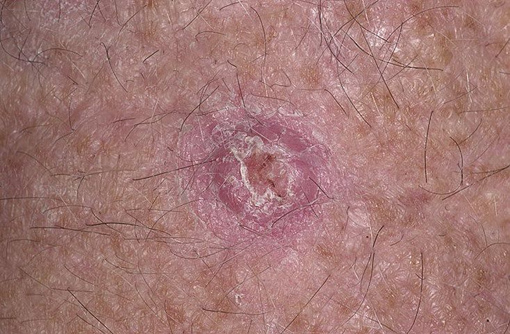
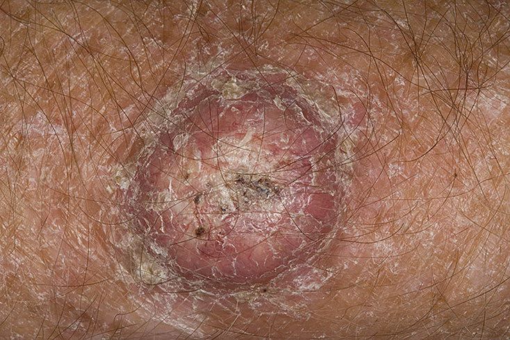
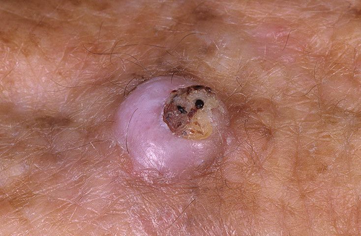
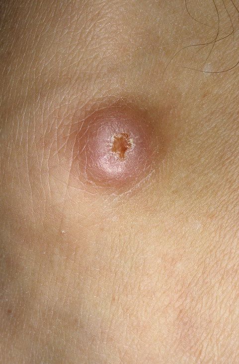
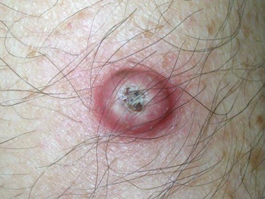
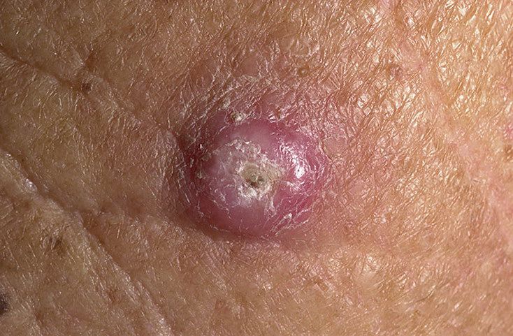
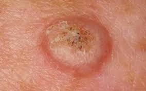
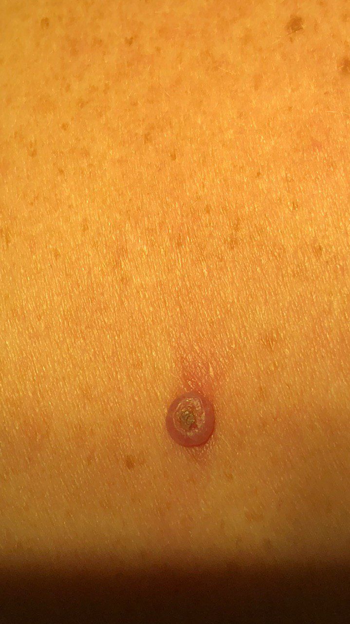
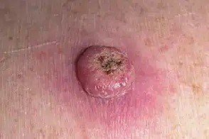
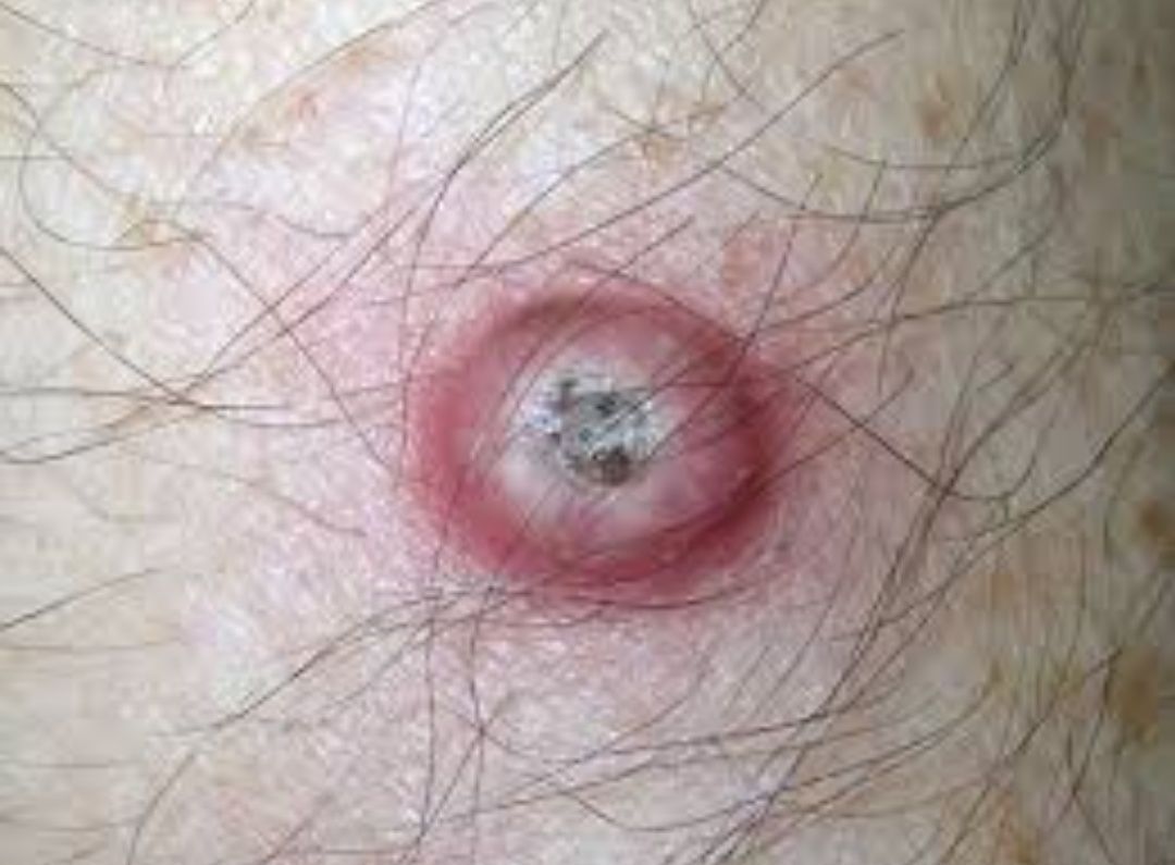
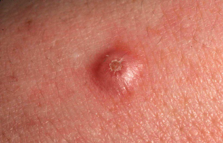
With a visual examination of keratoacanthomas, an elevated, flattened formation is determined. The surface at the edges is smooth, the skin pattern is absent, a crust-covered indentation, or, on the contrary, a protrusion formed by excess horn masses is noted in the center. In large tumors or when the stratum corneum disappears, ulceration may be present in the center.
The boundaries are usually fuzzy (although a clear border with healthy skin can sometimes be present), but they are even. The peripheral part of keratoacanthoma is usually represented by an epithelial roller. Around there may be a reaction of healthy skin in the form of hyperemia. The shape of the tumor is regular, symmetrical.
Coloring on the periphery is matte pink, pink-red, yellowish shades may be present. In the center, in the presence of horny masses – gray shades. When the crusts leave, a pink-red surface with possible foci of ulceration.
Hair growth is absent.
Sizes range from 4 mm to 40 mm. Swelling up to 20 mm is usually rapid. Above 20 mm, the keratoacanthoma grows slowly, but at this stage, soreness joins even with a slight physical impact, bleeding from the central part.
On palpation, a dense, but mobile formation with respect to the subcutaneous structures is determined.
Subjective sensations are usually absent. However, in formations over 15 mm there is an increased tactile sensitivity, soreness.
Keratoacanthoma is located mainly in open areas of the body. The most favorite localization is the forearm and the back surface of the hand. It can also be found on the face, neck, back, legs, in the chest area. A little less often – the area of the abdomen, hips.
Dermatoscopic Description
With dermatoscopy, the most reliable symptoms and signs of keratoacanthomas include:
- Homogeneous pink staining on the periphery;
- A whitish pink or white annular peripheral region;
- The accumulation of keratin masses in the center surrounded by a ring-shaped roller;
- Inclusions in the form of small blood clots (more characteristic for the central region);
- Peripheral vascular pattern;
- Vessels in the form of a hairpin;
- Linear vessels;
- The radial orientation of the vessels.
Differential diagnosis
Differential diagnosis is carried out with such neoplasms as:
- Skin horn;
- Dermatofibroma;
- Open comedone;
- Seborrheic keratosis;
- Bowen’s disease;
- Squamous cell carcinoma;
- Basal cell carcinoma;
- Melanoma.
Risks
Keratoacanthoma is an optional precancerous condition. Malignancy occurs rarely and is more often observed under the influence of additional factors (chronic injury, thermal and chemical burns). In the case of malignancy, keratoacanthoma transforms more often into squamous cell (squamous) cancer.
It should be taken into account that patients with keratoacanthoma have an increased risk of developing a malignant tumor on the unchanged skin or near the keratoacanthoma. This can complicate the timely detection and differential diagnosis of tumors.
Tactics
If keratoacanthomas are detected or suspected, an oncologist should be consulted. Due to the high degree of similarity of keratoacanthomas with basal cell carcinoma or squamous cell carcinoma, a thorough differential diagnosis should be carried out, including a morphological examination after a biopsy.
With histological confirmation of keratoacanthomas, despite the benign nature of the tumor and the possibility of its spontaneous involution, observation tactics are not recommended. This is due to the rapid growth of education, the appearance of soreness and bleeding when reaching large sizes (over 20 mm), as well as the risk of malignancy.
In case of refusal of the patient from surgical treatment, active dynamic observation is necessary. Moreover, photographic fixation of keratoacanthomas is of great value, which will make it possible to determine even minor changes in its appearance in the future.
Due to the increased risk of malignancy, as well as the appearance of other potentially dangerous skin neoplasms, a dermatologist or oncologist is examined in the spring and autumn (before and after the beach season). Of great importance is the mapping of skin neoplasms, which greatly simplifies further observation, the search for new formations, or changes to existing ones.
Treatment
The main method of treatment is surgical: excision of keratoacanthomas with the capture of healthy skin to the entire thickness. This is the most effective method with a low risk of local recurrence.
Excision along the plane is not recommended, since in this case, the likelihood of a reappearance of keratoacanthomas in the same place is high. Removal using local exposure methods (laser removal or cryodestruction) leads to similar consequences.
Prevention
Prevention of the appearance of keratoacanthomas is a gentle and careful attitude to the skin:
- Limitation of ultraviolet radiation (tanning bed, solar tanning);
- The use of protective creams during periods of active sun;
- Exclusion of chronic skin trauma;
- Limitation or exclusion of ionizing radiation, occupational hazards;
- Compliance with safety measures when working with skin-damaging factors;
- Personal hygiene and basic awareness of skin tumors.
It also requires regular examination of the skin, timely consultation with a specialist in the event of external changes in skin tumors, and the removal of potentially dangerous neoplasms.