Atopic dermatitis (syn: atopic eczema, endogenous eczema, diffuse neurodermatitis) is an acute, subacute or chronic inflammation of the epidermis and dermis, causing severe itching. A history or close relatives often have other allergic diseases (pollinosis, bronchial asthma, allergic rhinitis). Refers to the so-called atopic disease. In Greek, “atopy” means “without place”; in medicine, this concept combines a hereditary predisposition to atopic dermatitis, bronchial asthma and allergic rhinitis.
- In 60% of patients, the disease begins in the first year, most often in the third month of life.
- Men are somewhat more often affected in infancy and childhood, while in women it is more common in adolescence.
- 70% of patients have a history or close relatives with allergic rhinitis, pollinosis, bronchial asthma.
- In most patients, the disease begins before the age of 12 years, in adulthood the debut of the disease occurs very rarely.
- The course is chronic, relapsing, with periodic exacerbations.
- Seasonal exacerbations of allergies to airborne allergens (such as pollen) are noted.
Triggering factors
- Allergens: contact, air, food.
- Dry skin due to frequent washing of the body and hands.
- Emotional overload.
- Hormonal shifts: pregnancy, menstruation, thyroid disorders.
- Infections: Staphylococcus aureus, group A streptococci, dermatophytosis, candidiasis, herpes.
- Parasitic diseases (ascariasis, giardiasis, opisthorchiasis, enterobiasis, toxocariasis)
- Time of year: in temperate climates, there is an improvement in summer and an aggravation in winter.
- Clothing, especially woolen fabrics (itching increases after removal), woolen blankets, feather pillows.
Pathogenesis
Allergic reactions of the immediate type: interaction of the allergen with IgE-
by antibodies fixed on the surface of basophils and mast cells leads to the activation of these cells and the release of vasoactive substances. The role of IgE in the pathogenesis of atopic dermatitis is not entirely clear, but it has been established that Langerhans cells (specialized process cells of the epidermis) carry high-affinity IgE receptors and appear to mediate an inflammatory skin response.
Symptoms
Subjective
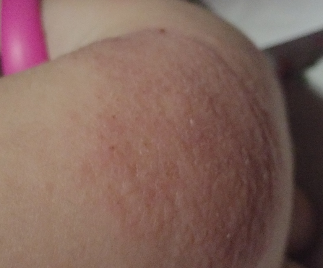
- Dry skin.
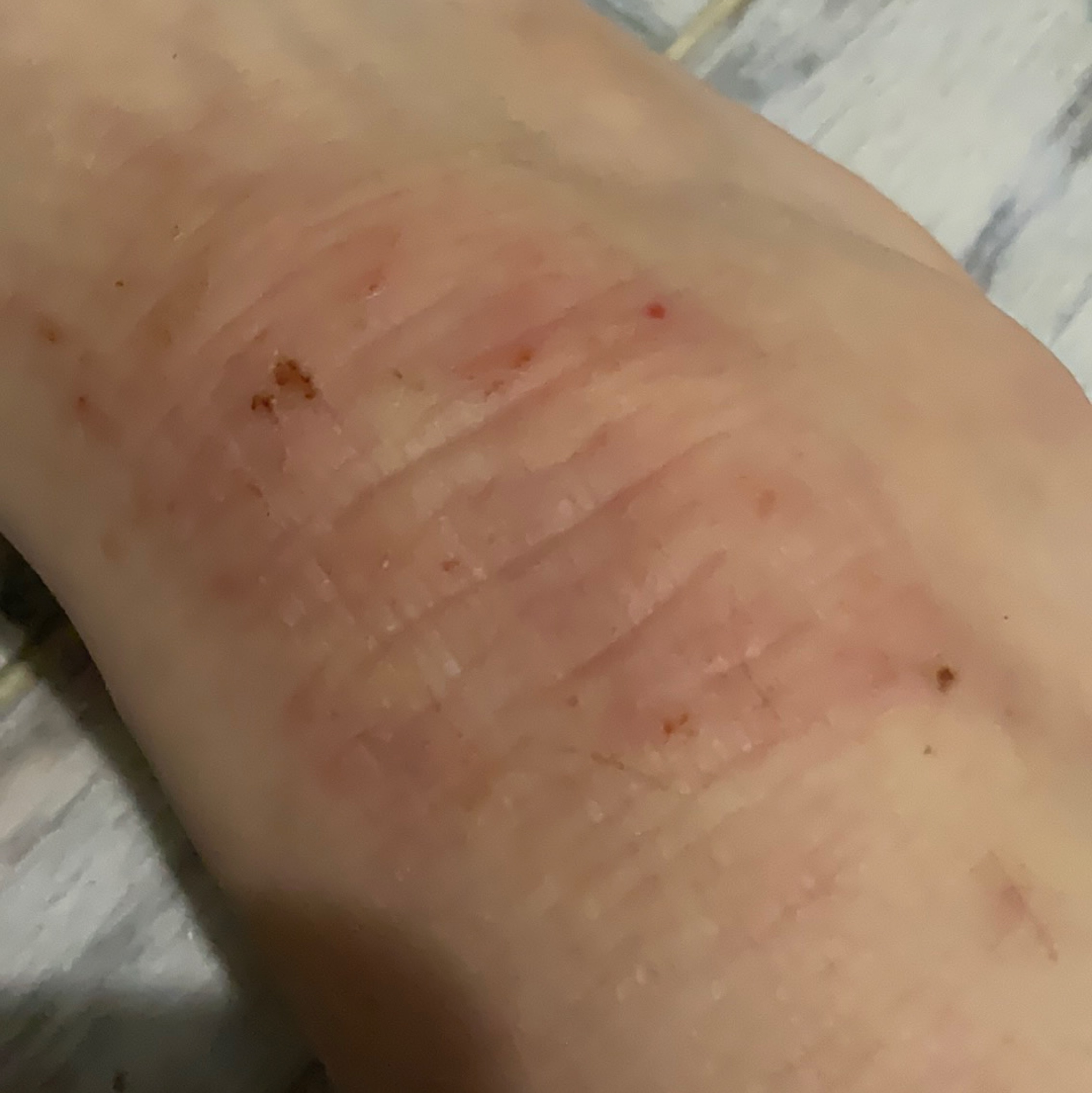
- The main symptom is skin itching. Constant scratching creates a vicious circle: itching -> scratching -> rash -> itching.
- Lichenization gradually develops.
Objective
Typical localization:
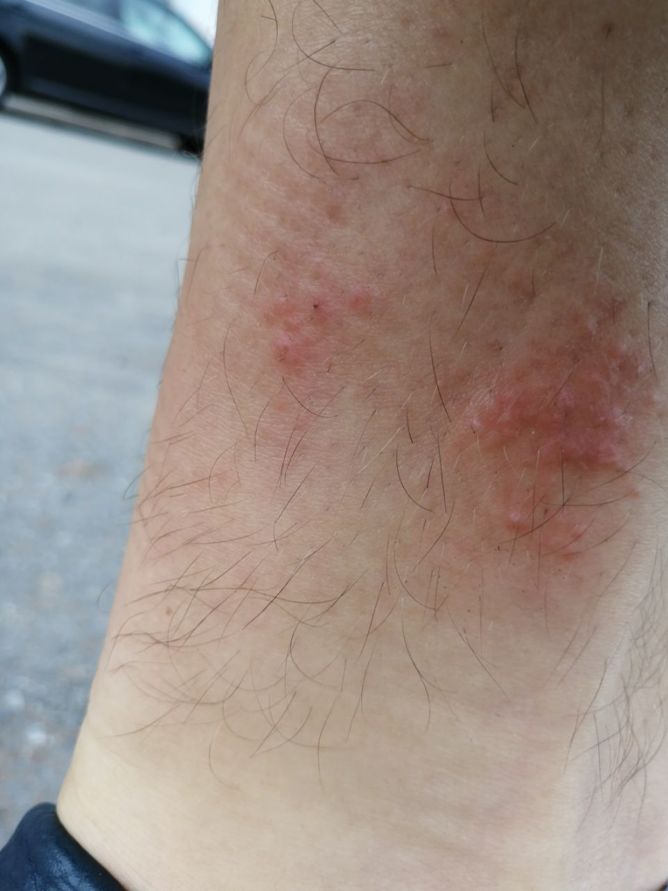
- flexor surfaces of the joints,
- the anterior and lateral surfaces of the neck,
- face (eyelids, forehead),
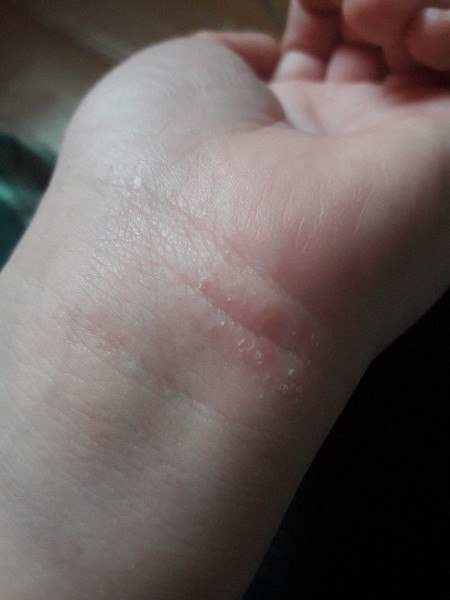
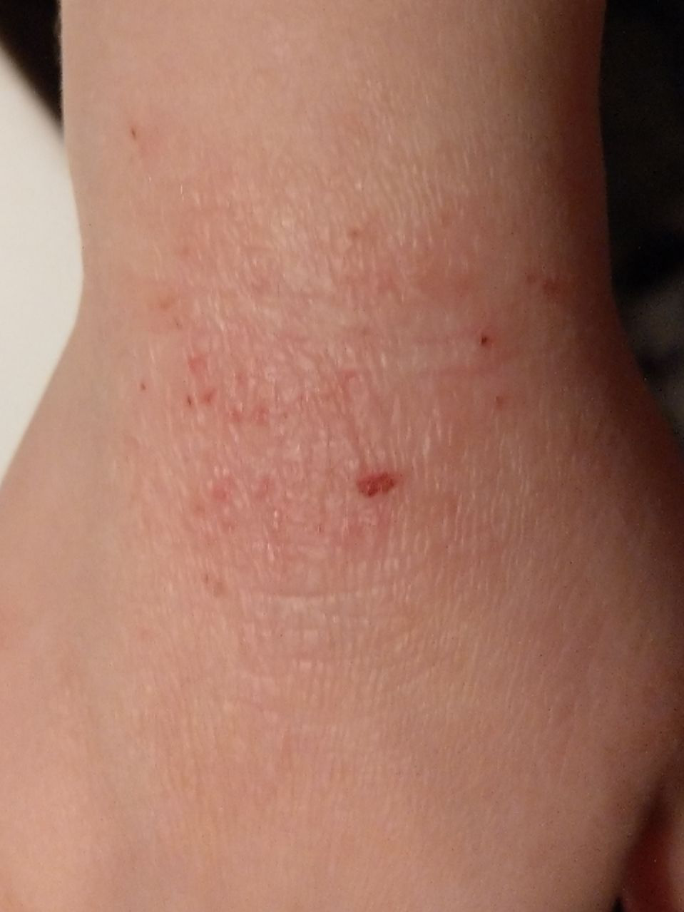
- wrists,
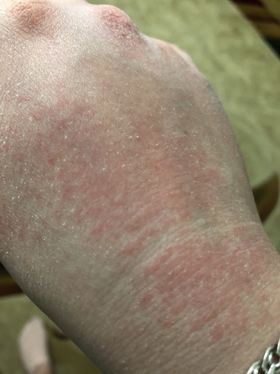
- the back of the hands and feet,
- In severe cases, a generalized lesion.
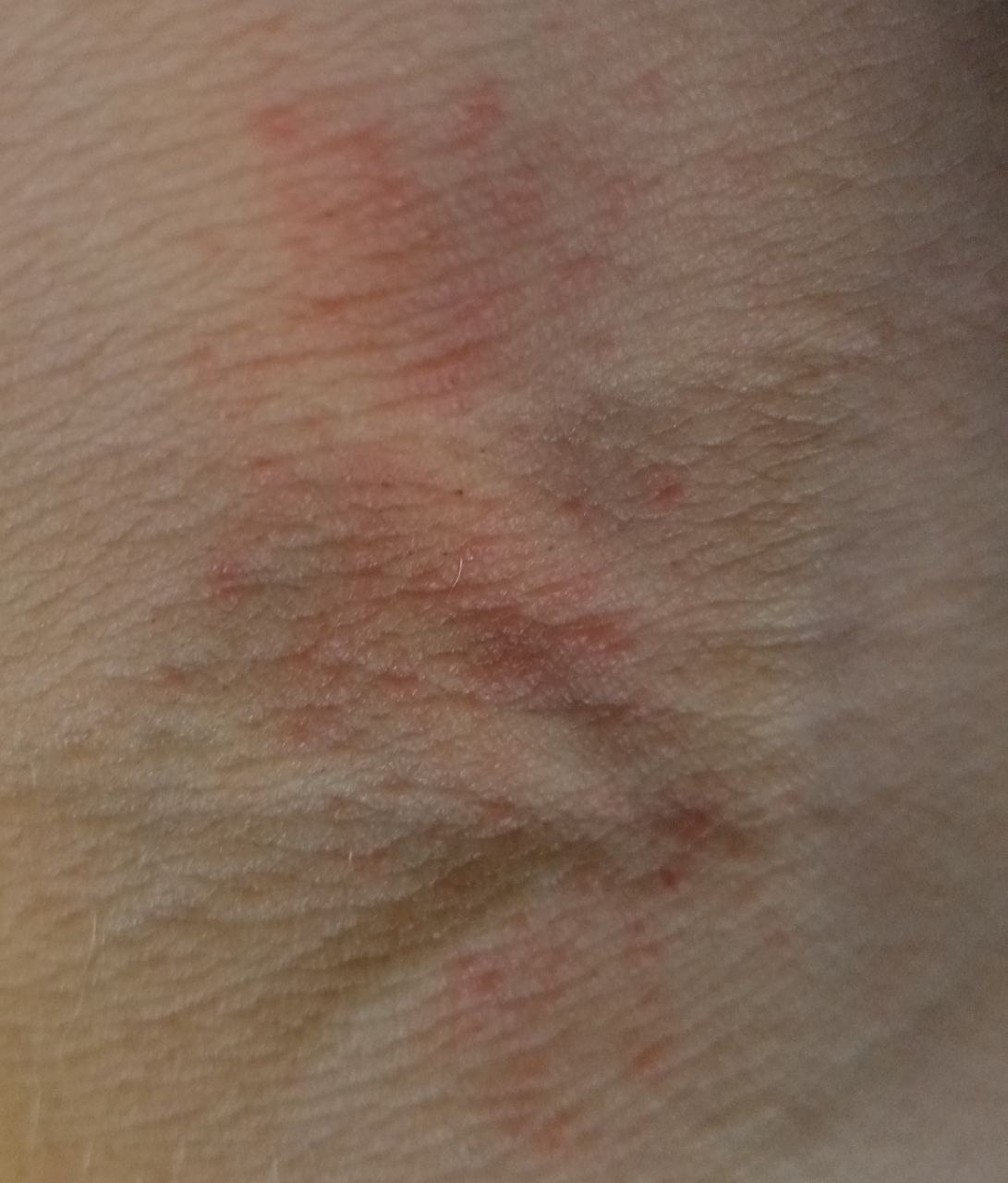
Elements of the rash
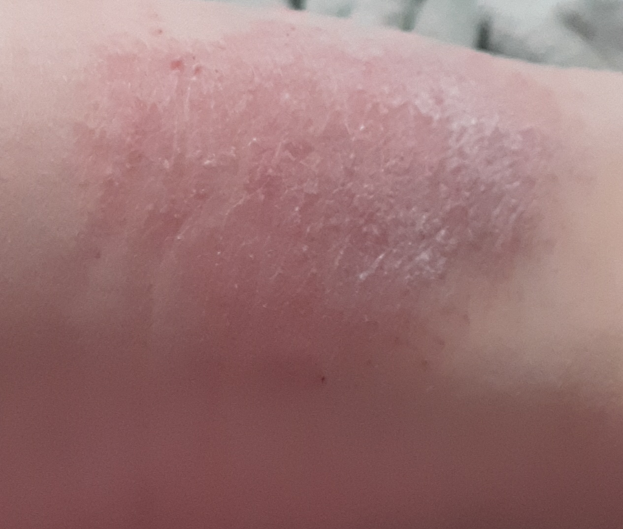
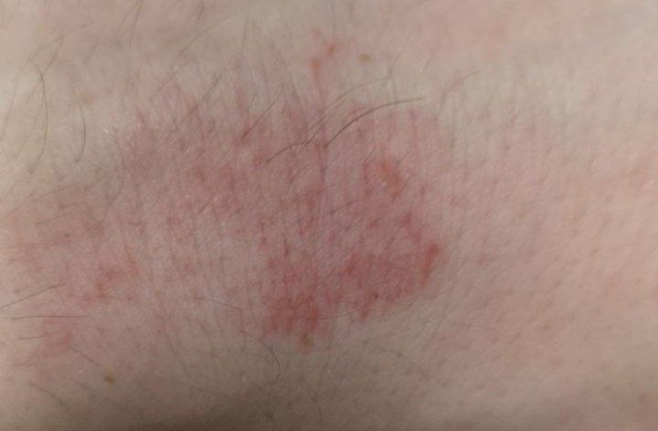
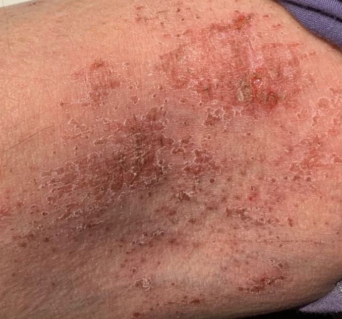
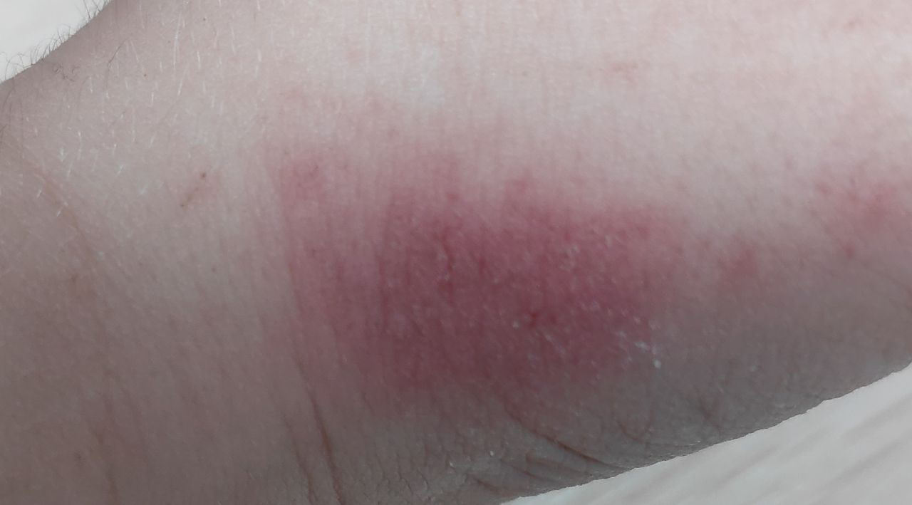
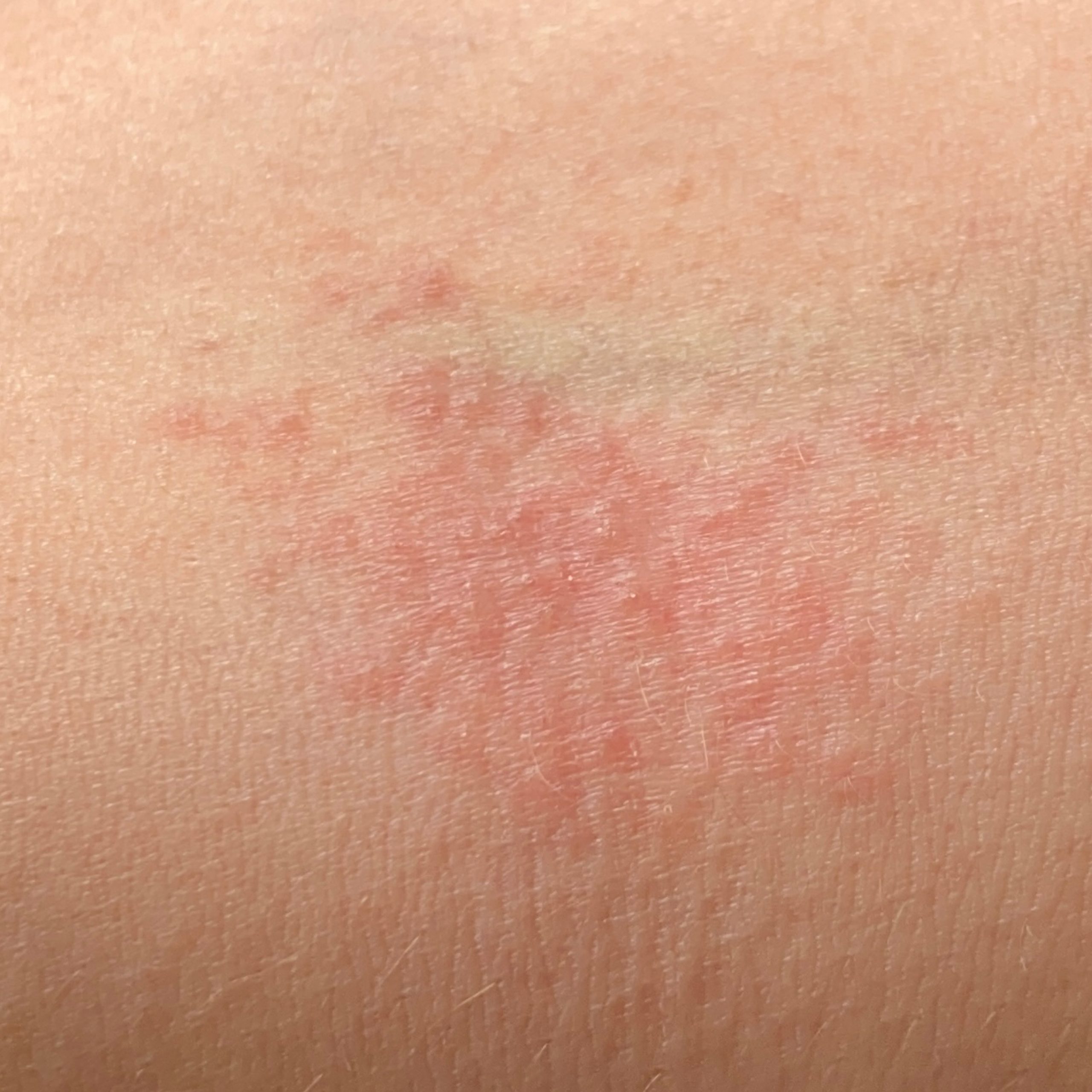
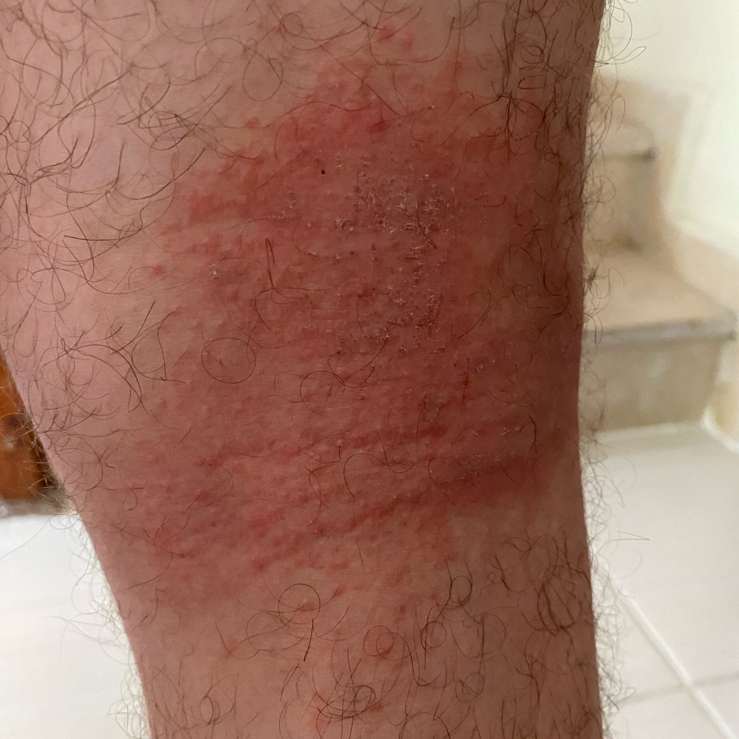
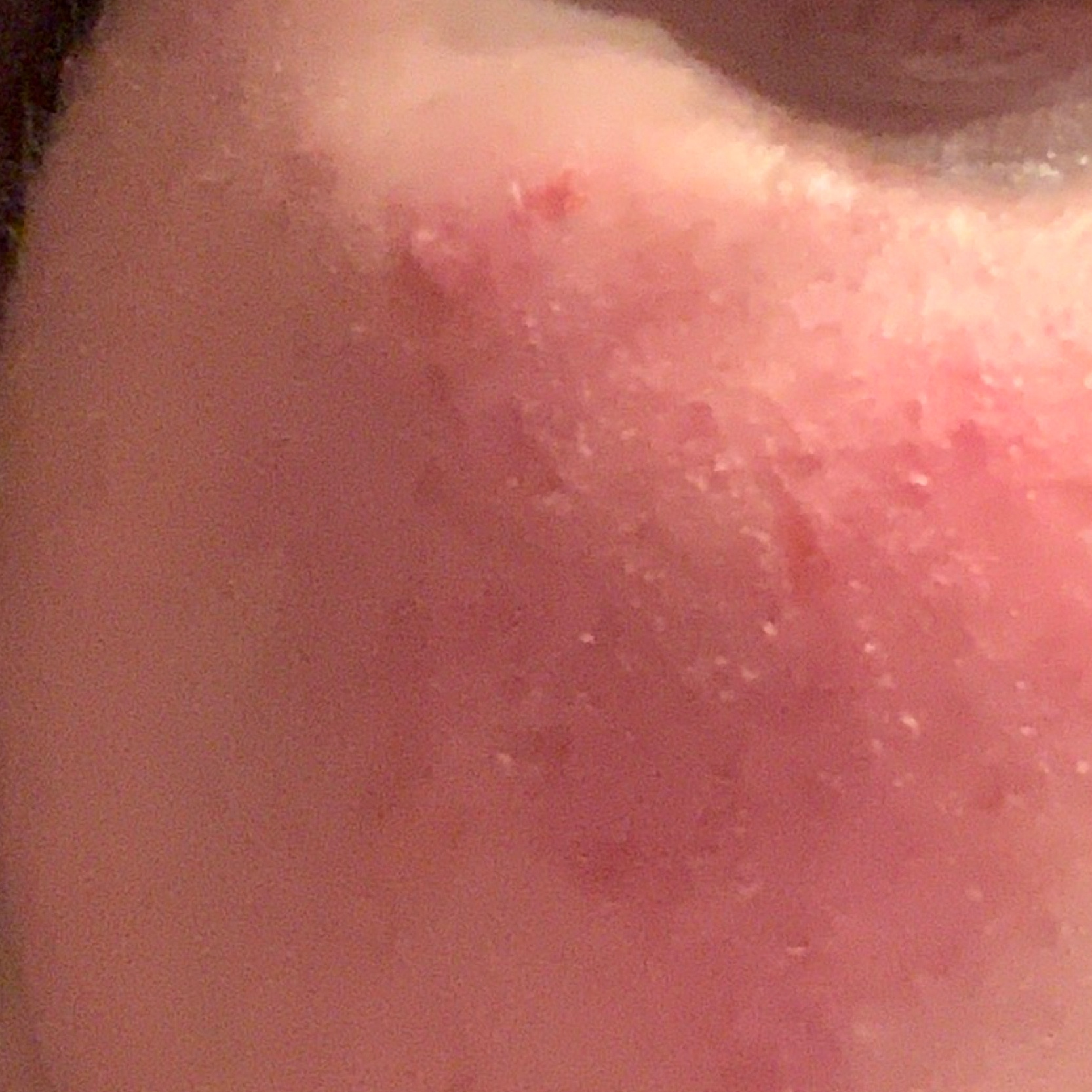
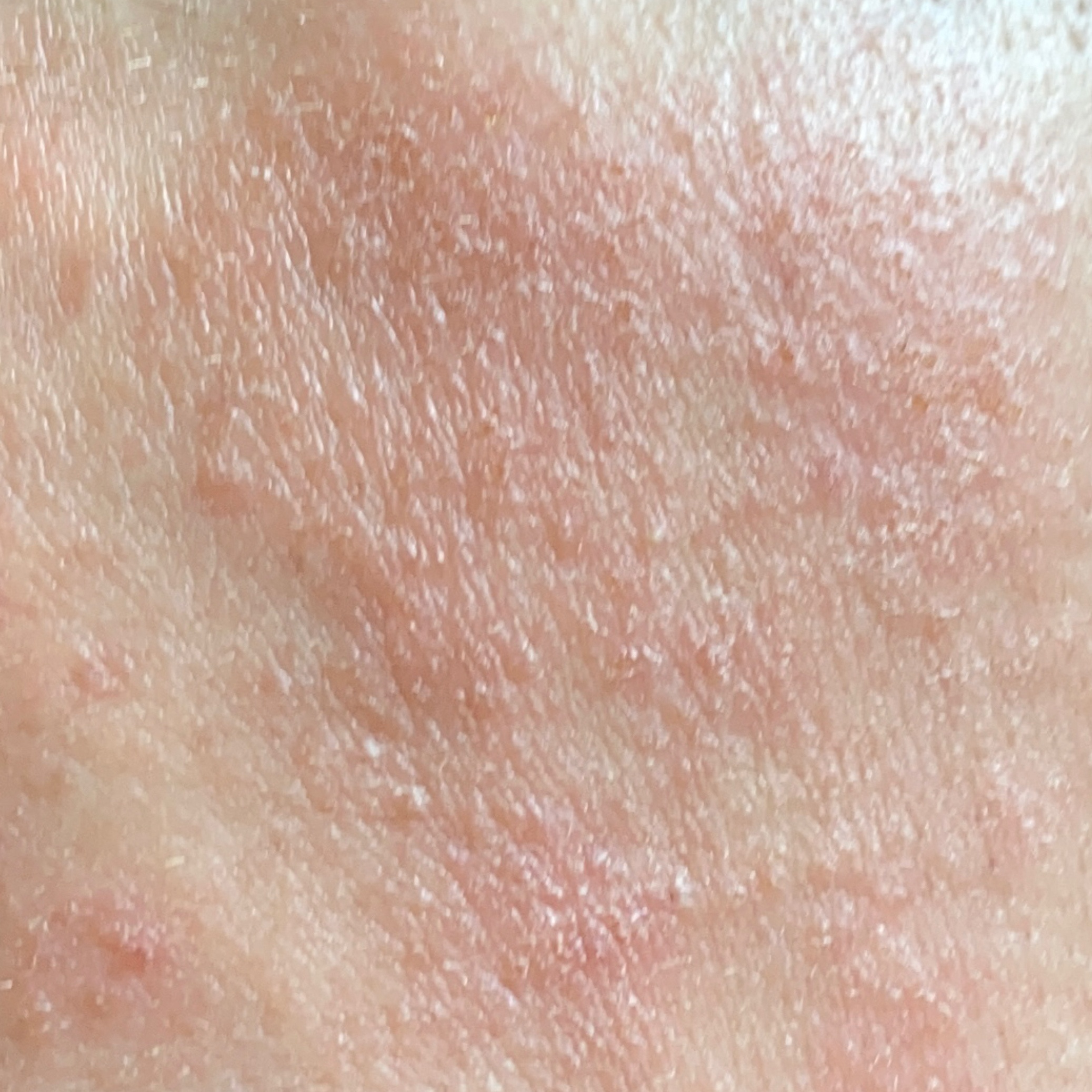
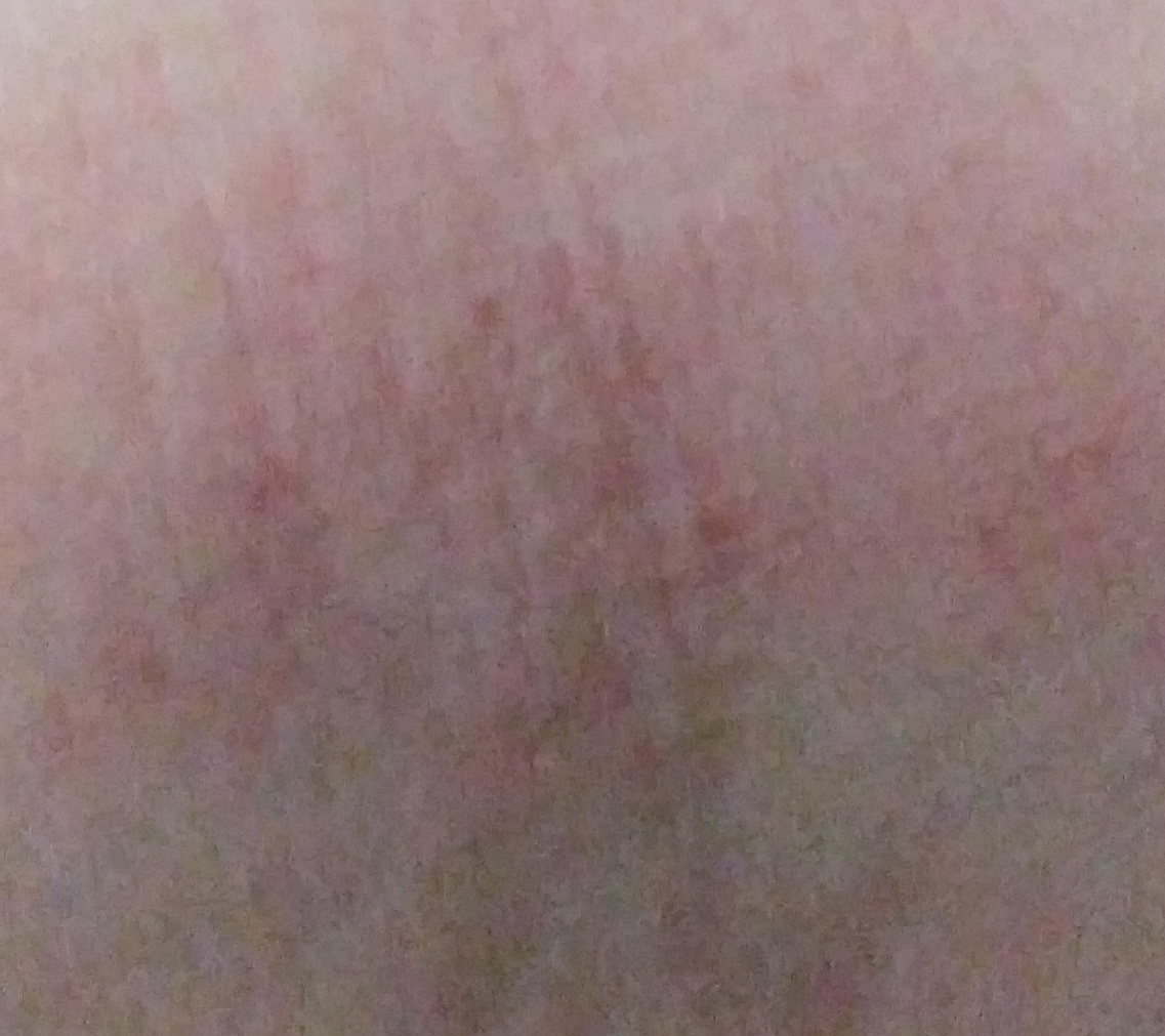
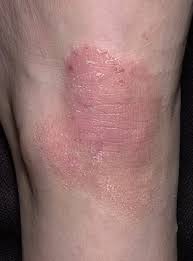
The acute stage.
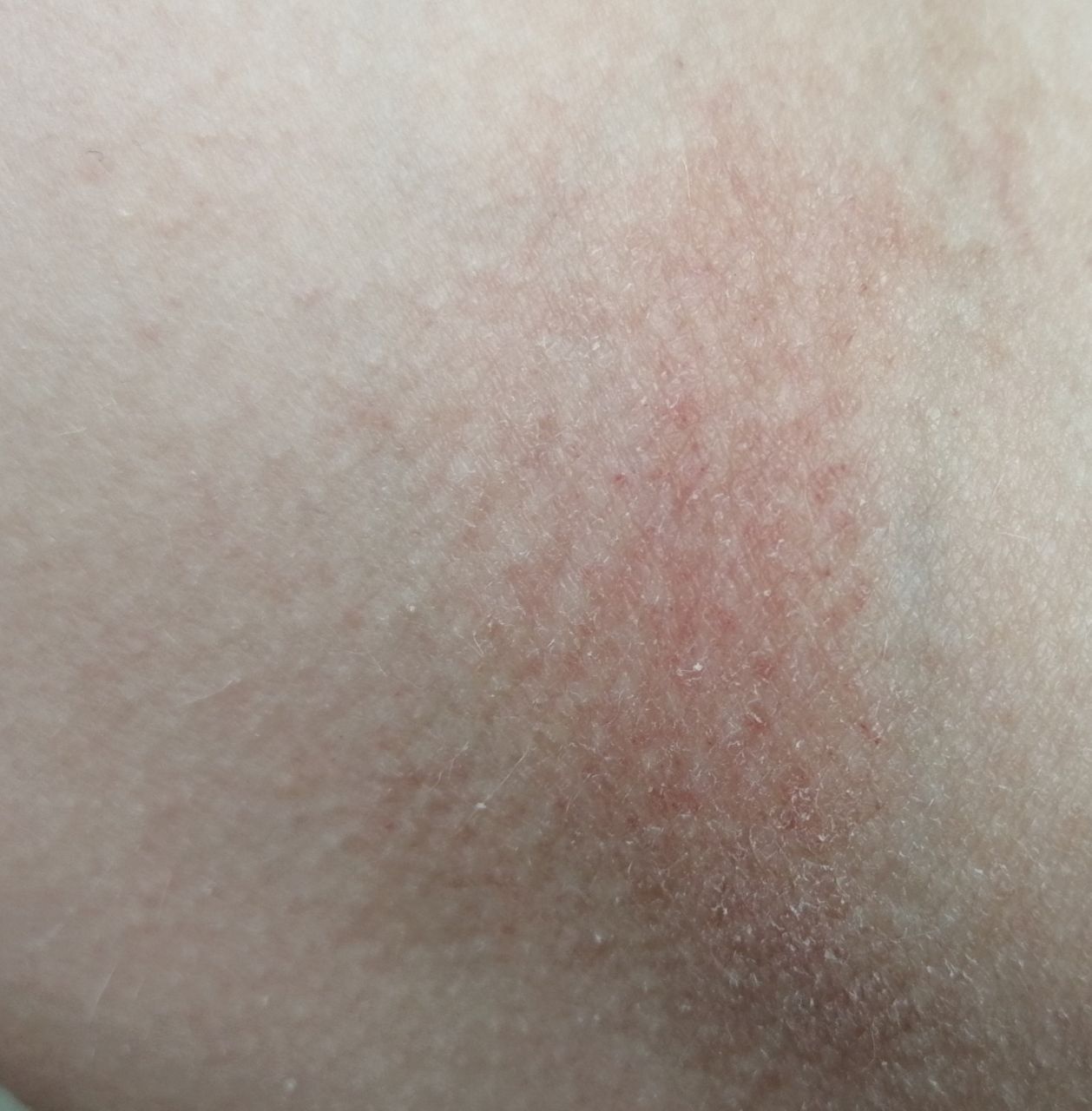
- Red spots with fuzzy borders, papules and plaques, often peeling.
- Widespread swelling (the skin looks puffy, puffy).
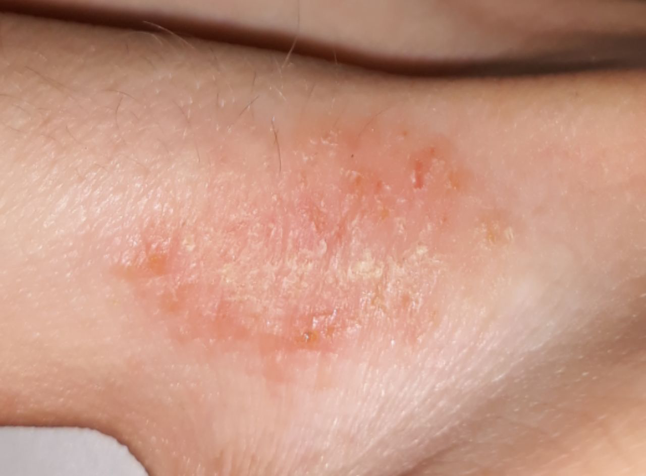
- Erosion, blotting, and crusts.
- Excoriations are the result of scratching.
- Secondary infections caused by Staphylococcus aureus.
- Pustules (usually pierced in the center by a hair).
- Corky.
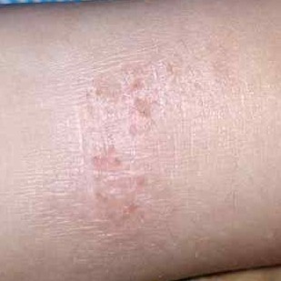
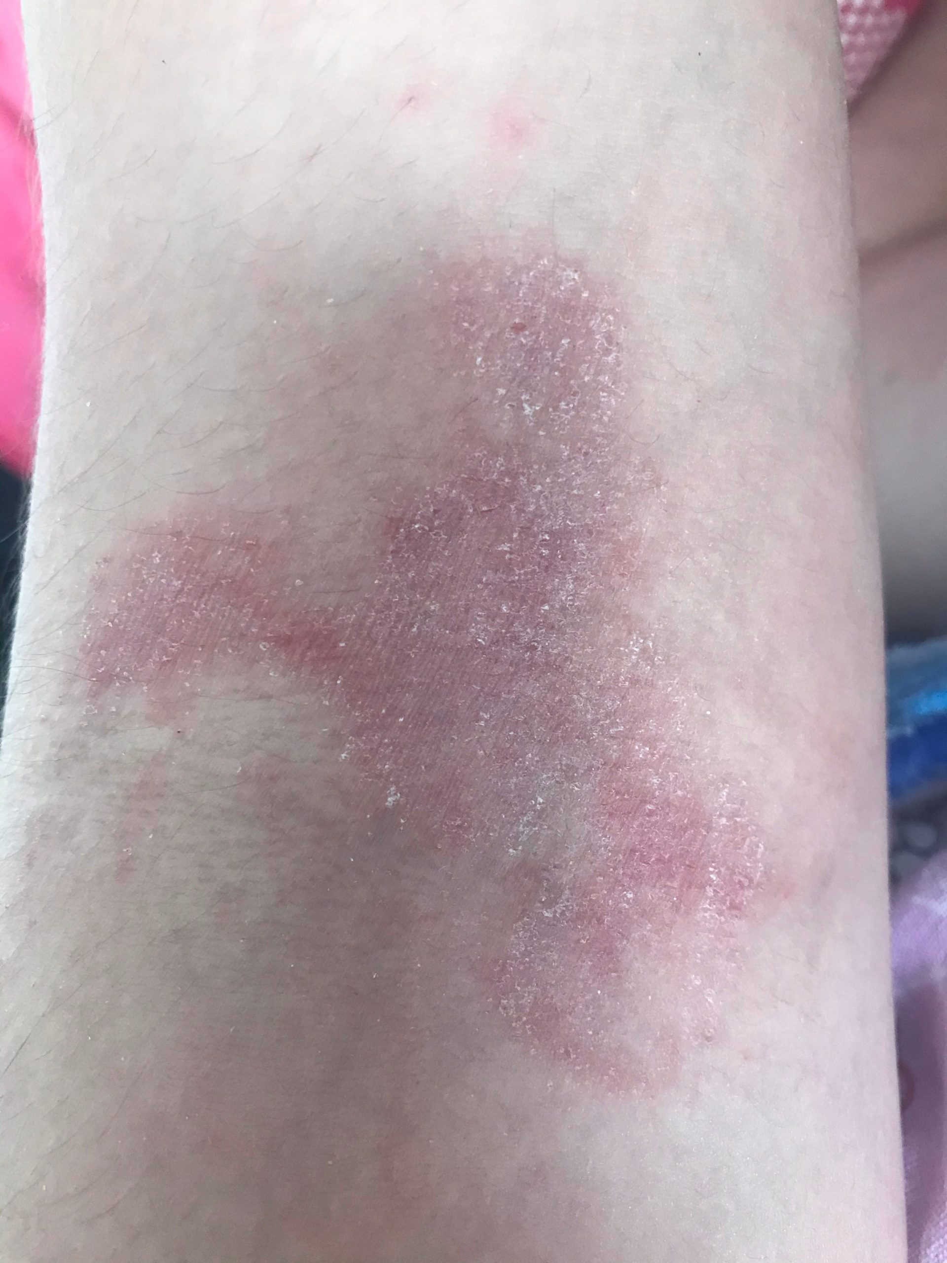
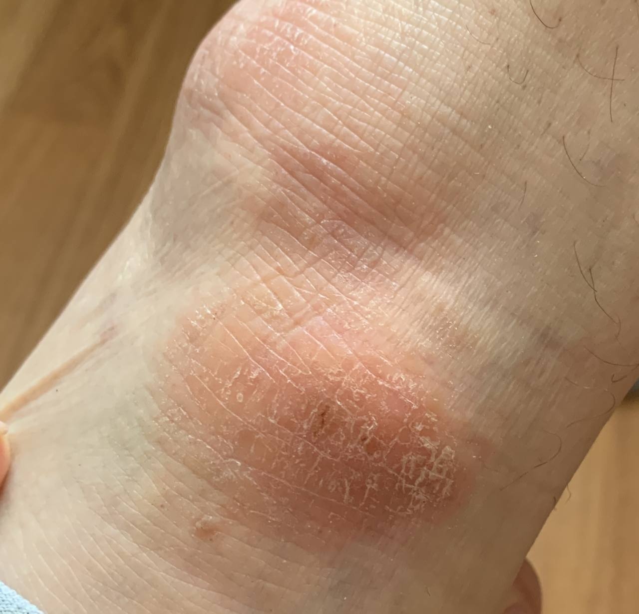
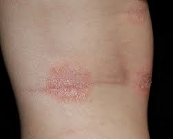
Chronic stage.
- Lichenization (thickening of the skin and increase of the skin pattern) is the result of constant scratching and rubbing.
- Many small papules on the site of hair follicles.
- Cracks are painful, especially on the palms, fingers and soles.
- Loss of the outer third of the eyebrows and hyperpigmentation of the eyelids are the result of scratching the eyes.
- Characteristic skin fold under the lower eyelid (Denny-Morgan lines) .
- White dermographism is a very characteristic sign of atopic dermatitis: if you run a blunt object over the affected skin, after a few seconds because of capillary spasm, a white stripe appears.
- Common ichthyosis and follicular keratosis are detected in 10% of patients.
Peculiarities of atopic dermatitis in infants
In infants, atopic dermatitis is one of the most severe skin diseases.
As a rule, there is a hereditary predisposition to allergic diseases. The most common allergens are food products.
Elements of the rash.
Skin is red and edematous, covered with vesicles; peeling, mottling, crusts, cracks.
Localization.
Individual areas of the body are affected. Typical localization is face (except lips) and extensor surfaces of extremities.
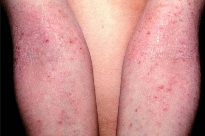
Peculiarities of atopic dermatitis in older children
Elements of the rash.
Papules and plaques, lichenization, erosions, crusts.
Localization.
The most common are the ulnar fossa and the popliteal fossa.
Features of atopic dermatitis in adults
Atopic dermatitis in adults is a chronic disease with exacerbations,
which are often caused by emotional overload. Many patients suffered from atopic dermatitis in childhood, often accompanied by bronchial asthma, allergic rhinitis.
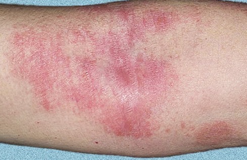
Elements of the rash
Acute stage. Red plaques with fuzzy borders; often peeling. Excoriations appear due to scratching of skin. With secondary infection, pustules (usually pierced by hair in the center) and crusts appear.
Chronic stage. Lichenization is a thickening of the skin with an intensification of the skin pattern, which appears due to constant scratching of the skin. Lichenization foci cover large areas of the skin or represent individual nodules (which can easily be confused with nodular scabies).
Aggravation. Papules and plaques, lichenization, exoriations, pustules, erosions, dry crusts or pustules, cracks.
Localization.
Generalized lesions are common. Typical localization: flexural surfaces of arms and legs, front and side surfaces of the neck, face (eyelids, forehead), wrists, rear surface of feet and hands.
Course and prognosis
As the child grows older, there is usually a more or less stable remission. For adolescents, exacerbations, which are more severe, are characteristic. Most have the disease for 15-20 years. In 30-50% of them develop bronchial asthma or pollinosis. If atopic dermatitis begins in adulthood, it is very severe.
Complications
Secondary staphylococcal infection results in extensive erosions and crusts. Kaposi’s herpetic eczema is a very severe, sometimes life-threatening complication caused by the herpes simplex virus.
Differential diagnosis
- seborrheic dermatitis,
- simple and allergic contact dermatitis,
- psoriasis,
- coinous eczema,
- dermatophytosis,
- skin lymphoma (early stages).
- enteropathic acrodermatitis,
- celiac disease,
- glucagonoma,
- histidinemia,
- phenylketonuria;
- diseases of the immune system – Wiskott-Aldrich syndrome, X-linked agammaglobulinemia, IgE hyperproduction syndrome, histiocytosis X (Letterer-Sivet syndrome), isolated IgA deficiency.
Diagnosis
Anamnesis (onset in infancy) and clinical picture (typical localization and elements of rash, white dermographism).
Additional methods of examination
- Seeding: the nasal cavity and affected skin are often infected with Staphylococcus aureus. In the severe form of atopic dermatitis, carrier or secondary Staphylococcus aureus infection is detected in almost 90% of patients.
- Virus isolation in cell culture: if crusts are present, Kaposi’s herpetic eczema (the causative agent is herpes simplex virus) is ruled out.
- Determination of total IgE concentration in blood
- Skin tests with allergens (prick test, scarification tests, intradermal tests).
- Food allergen provocation tests.
- Histological examination in doubtful cases: the epidermis contains acanthosis, expressed in varying degrees, occasionally spongiosis (intercellular edema). Dermal infiltrates contain lymphocytes, monocytes, mast cells; few or no eosinophils.
- Radioimmunosorbent test for IgE
Treatment
- Hypoallergenic diet
- Control of the patient’s environment
- External therapy and skin care
- Systemic pharmacotherapy
- Treatment of combined allergic diseases (bronchial asthma, allergic rhinitis)
- Treatment of detected systemic and organ manifestations of atopic dermatitis (pathology of the gastrointestinal tract, urinary system, thyroid gland)
- Rehabilitation treatment
- Psychotherapy
- Educational programs for family members and patients