Melanoma is an aggressive malignant tumor. It develops from melanocytes (cells containing the pigment melanin). Due to the fact that this type of cell is found not only on the skin but on the mucous membranes and in the retina – melanoma can appear in other anatomical areas (eyes, genitals, rectum, soft tissues). About 95% of all melanomas are located on the skin.
The aggressiveness of melanoma is determined by the ability of this tumor to frequent relapses and metastasis in almost all organs. The metastasis pathway can be either lymphogenous or hematogenous. The rapid progression of melanoma also depends on the state of the body’s natural antitumor immunity.
A number of basic forms of melanoma are described:
- Superficially spreading: more typical for women, the prognosis is often favorable due to the small depth of invasion of the skin (70%);
- Nodular: more typical for men, the prognosis is often unfavorable due to the large depth of invasion of the skin (15%);
- Acrolentiginous or subungual melanoma: more typical for dark-skinned individuals. In addition to the nail bed, it is located at the fingertips and palms (10%);
- Lentiginous melanoma: more characteristic of women, appears against the background of age spots of lentigo, melanosis. The prognosis is often favorable due to the small depth of invasion of the skin (5%);
- Pigmentless melanoma: very rare.
Most often, melanoma appears in middle age (30-50 years). At an earlier age is extremely rare. For the older age group, lentiological forms of melanoma are especially characteristic.
Various factors lead to the transformation of normal melanocytic cells into malignant melanoma cells.
Factors that increase the risk of melanoma
There are a number of factors that, to varying degrees, can increase the risk of melanoma, although it is not appropriate to talk about any specific and unambiguous reasons for the appearance of a tumor:
- Natural (sun) and artificial ultraviolet;
- White / fair skin (I-II phototypes of the skin), the presence of pink freckles;
- Blue, gray or green eyes;
- Blond, red hair;
- Frequent sunburn, including a medical history (especially dangerous sunburn up to 14 years old);
- The presence of dysplastic melanocytic nevus, atypical nevus, blue nevus (especially multiple and congenital), Dubreuilh melanosis;
- A burdened hereditary history of melanoma (genetic factor);
- Pigment xeroderma;
- Previous melanoma;
- Age over 50;
- Sporadic or chronic injuries of the pigmented nevi (especially in traumatic places: collar, cuffs, belt, skin folds).
Diagnostics
Diagnosis of melanoma is based on a clinical examination, which includes a routine examination of the formation and dermatoscopy. The final decision in the diagnosis is only the result of histological examination. In addition to examining the tumor itself, diagnostics of regional and distant metastasis zones is carried out.
Symptoms
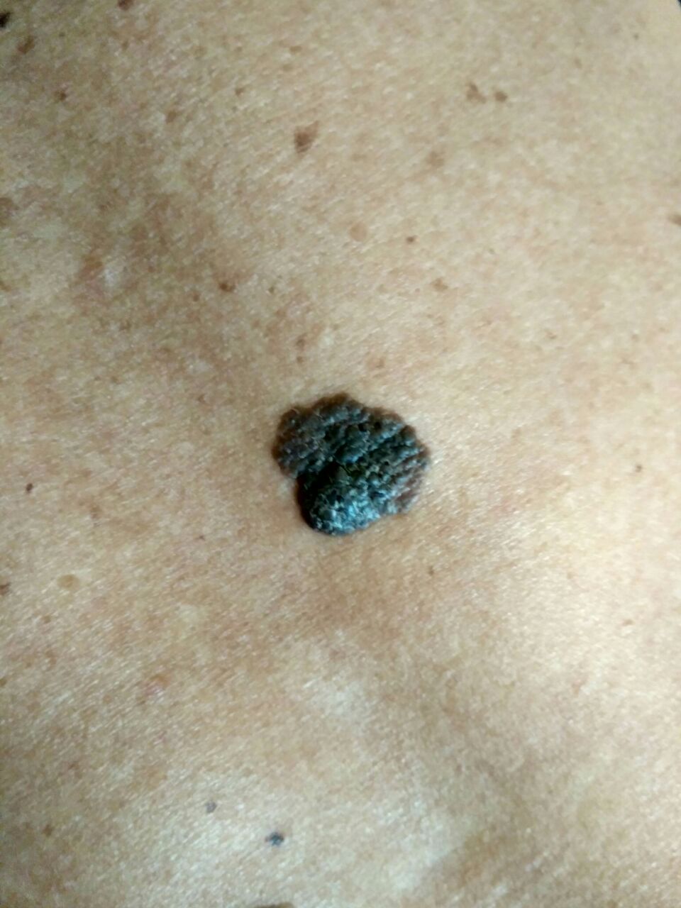
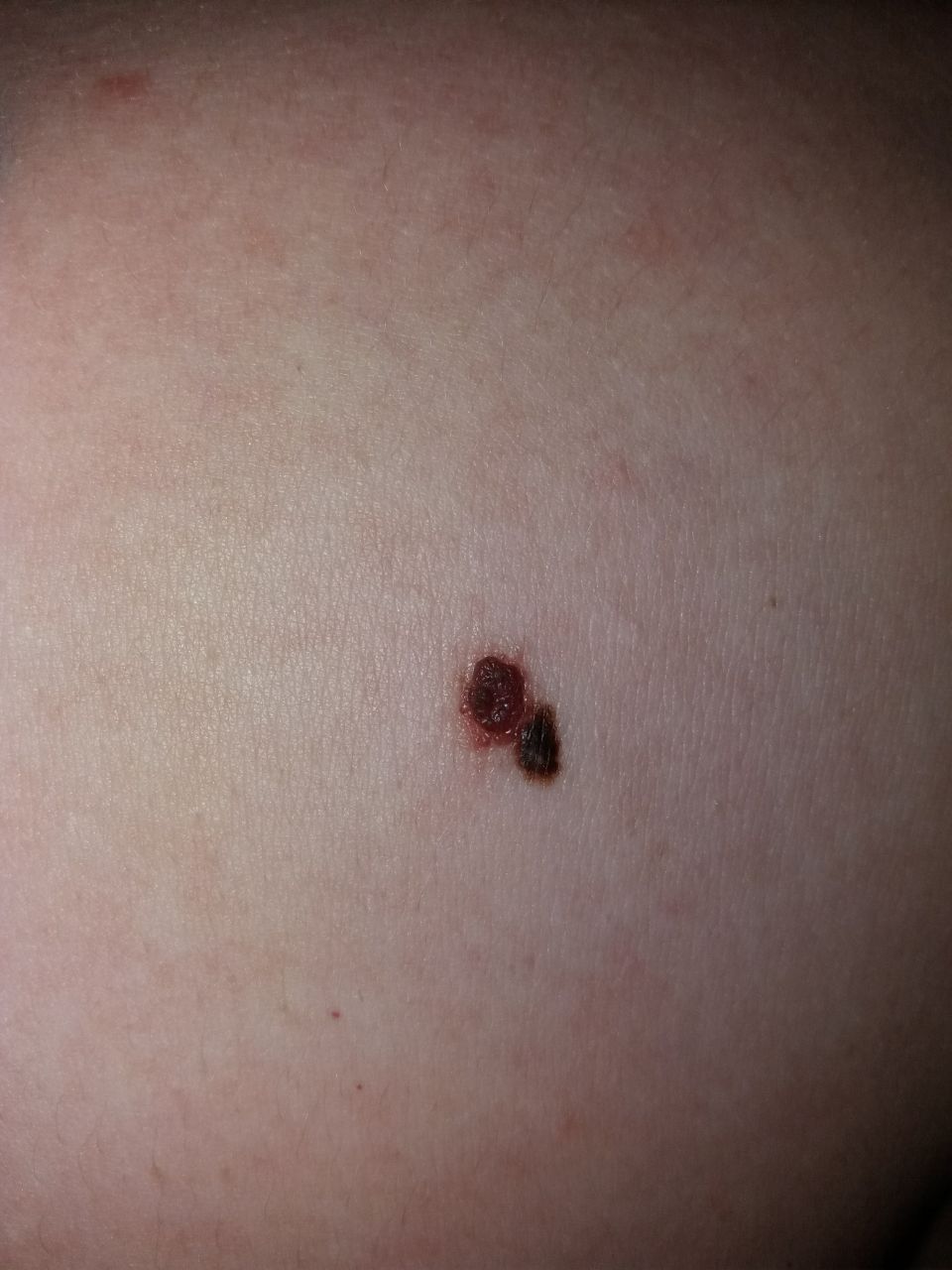
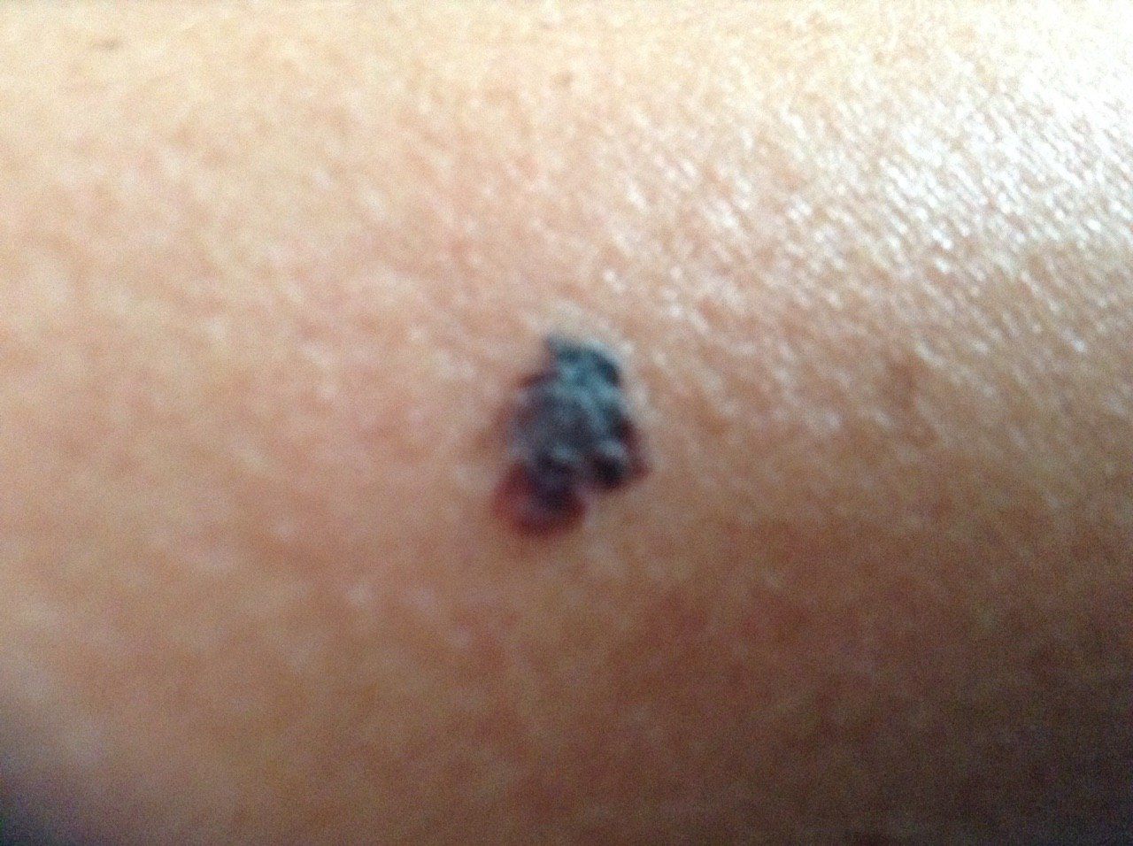
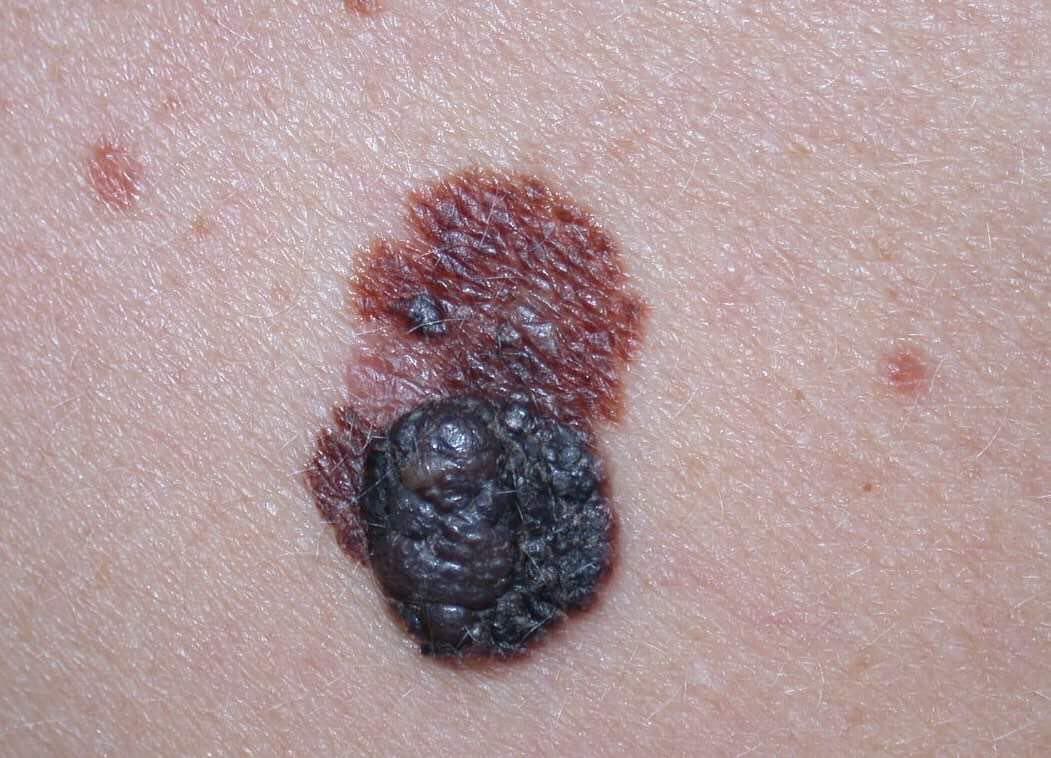
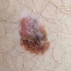
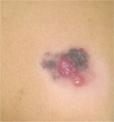
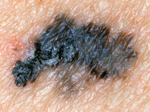
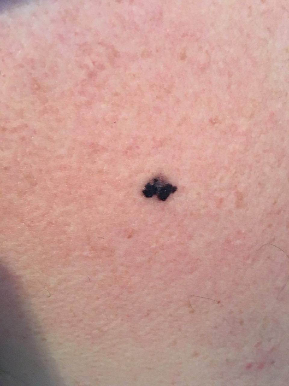
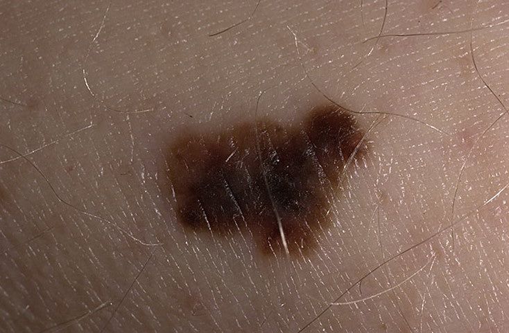
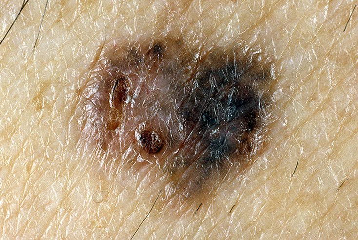
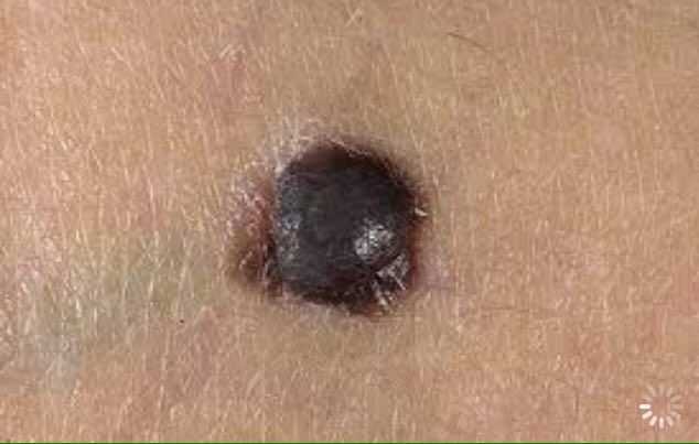
A visual examination of melanoma determines a multiform formation in the form of a spot, a tumor that rises above the skin, or a combination of these forms is noted. The surface of melanoma is usually different from the texture of ordinary skin. Only in the very early stages (zero or first) the skin pattern can not be disturbed. In other cases, there is smoothness, tuberosity, the presence of an exophytic component, weeping, ulceration, and bleeding.
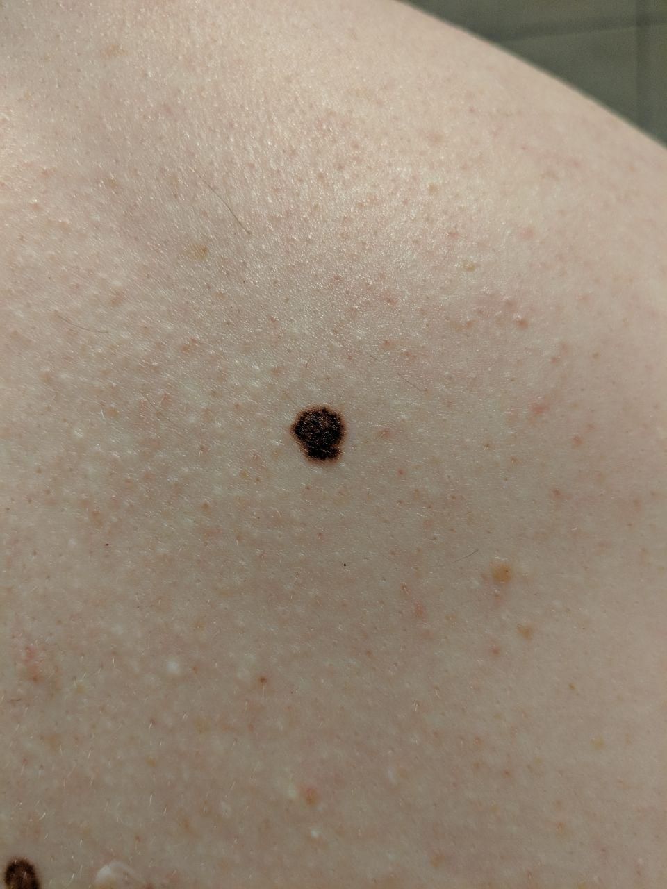
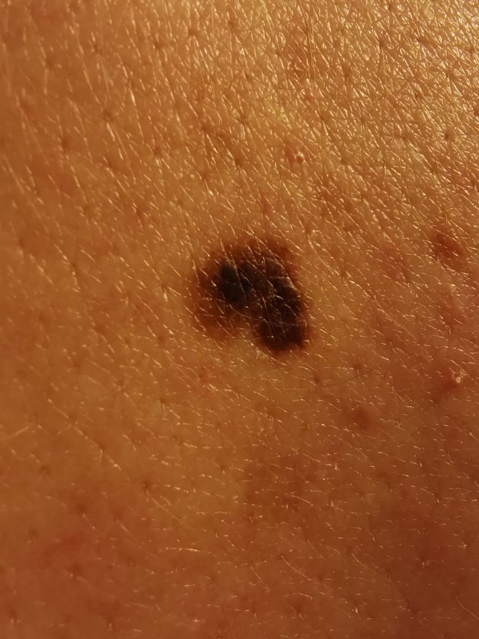
Visual assessment of pigmented lesions suspicious of melanoma is primarily carried out using the ABCDE system (Friedman, 1985):
A – asymmetry, asymmetry of the tumor;
B – border, the state of the edge of the tumor;
C – color, tumor pigmentation;
D – diameter, tumor size;
E – evolving, the evolution (change) of a tumor over time.
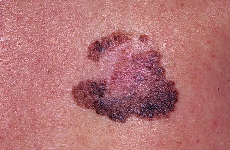
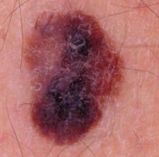
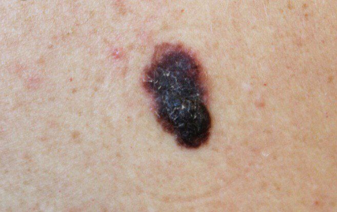
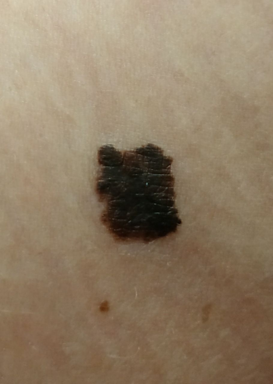
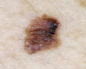
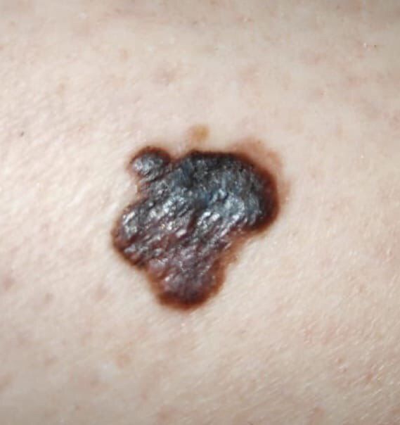
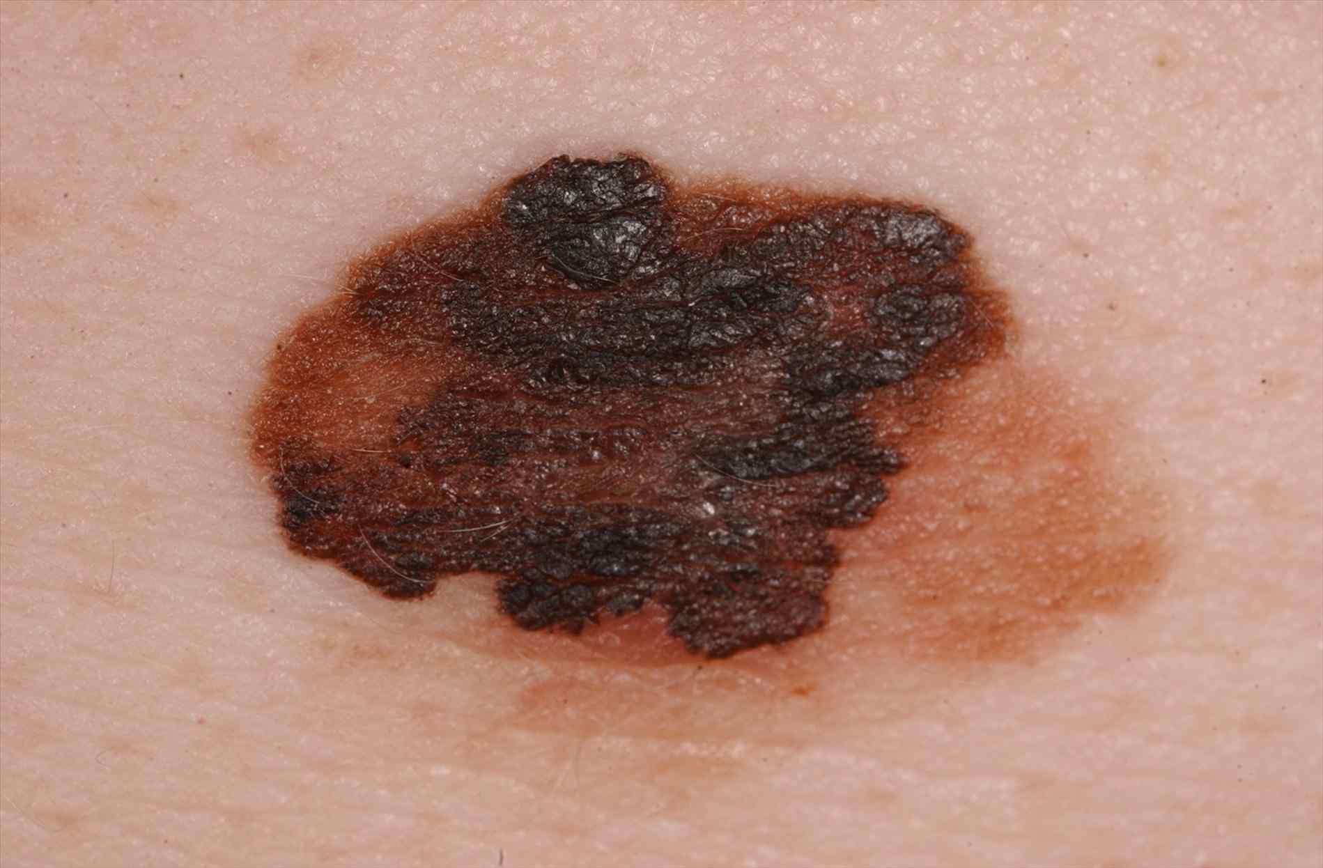
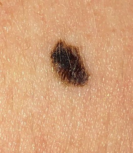
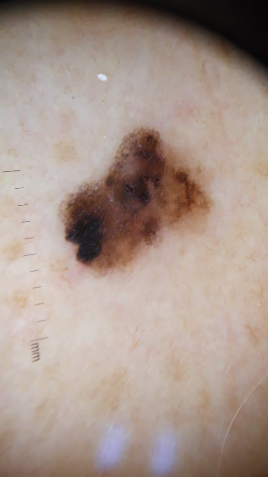
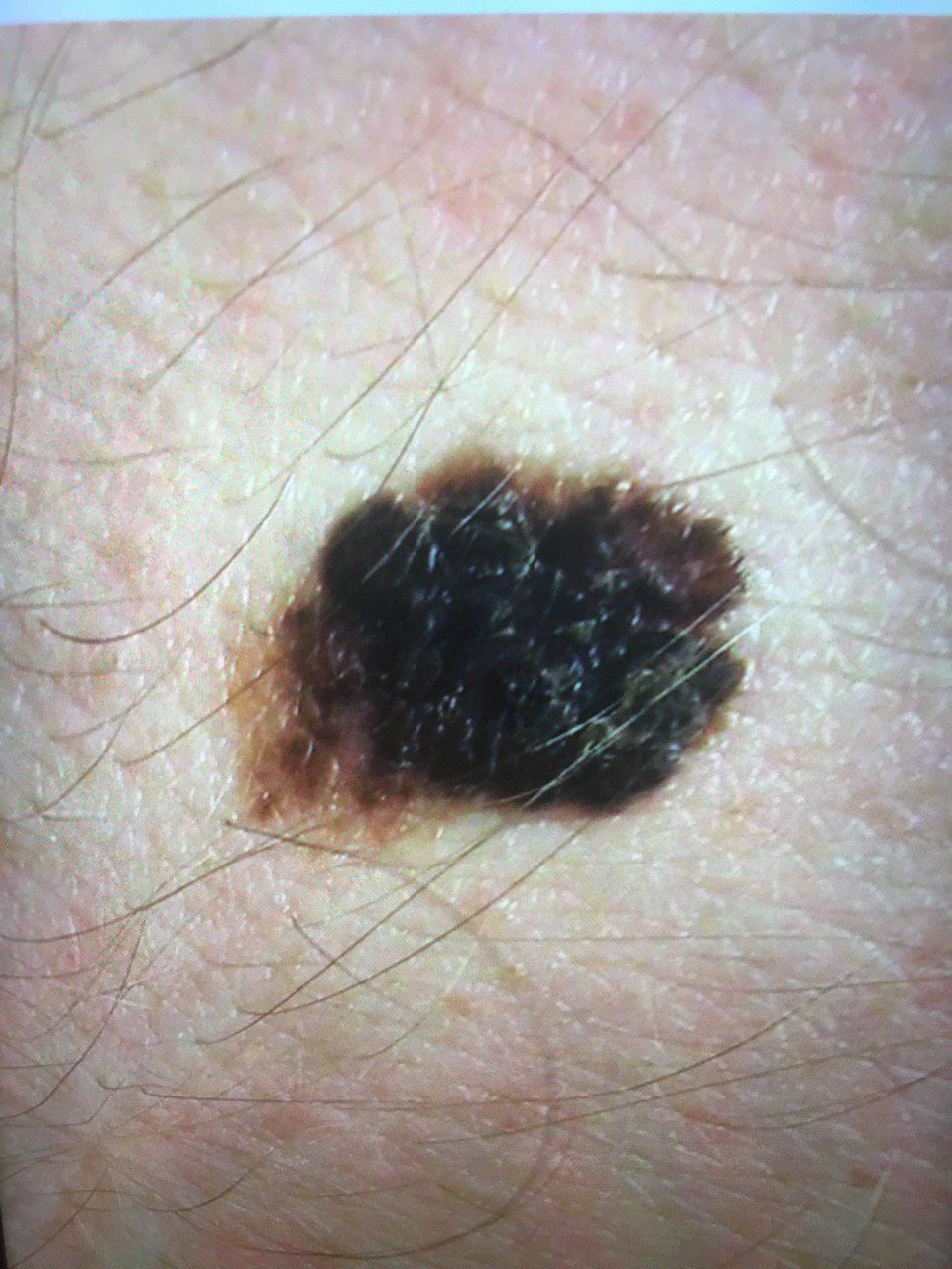
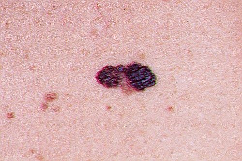
Asymmetry is an irregular form of a pigmented neoplasm. It is determined by drawing a conditional line through the center of the tumor, while one half will not be a mirror image of the other half.
The edges of melanoma (the border with healthy skin) are uneven. In surface forms, the edge is usually clear (favorable factor), when an invasion appears (vertical growth, “immersion” in the skin), the edge becomes less clear, blurred (unfavorable factor).
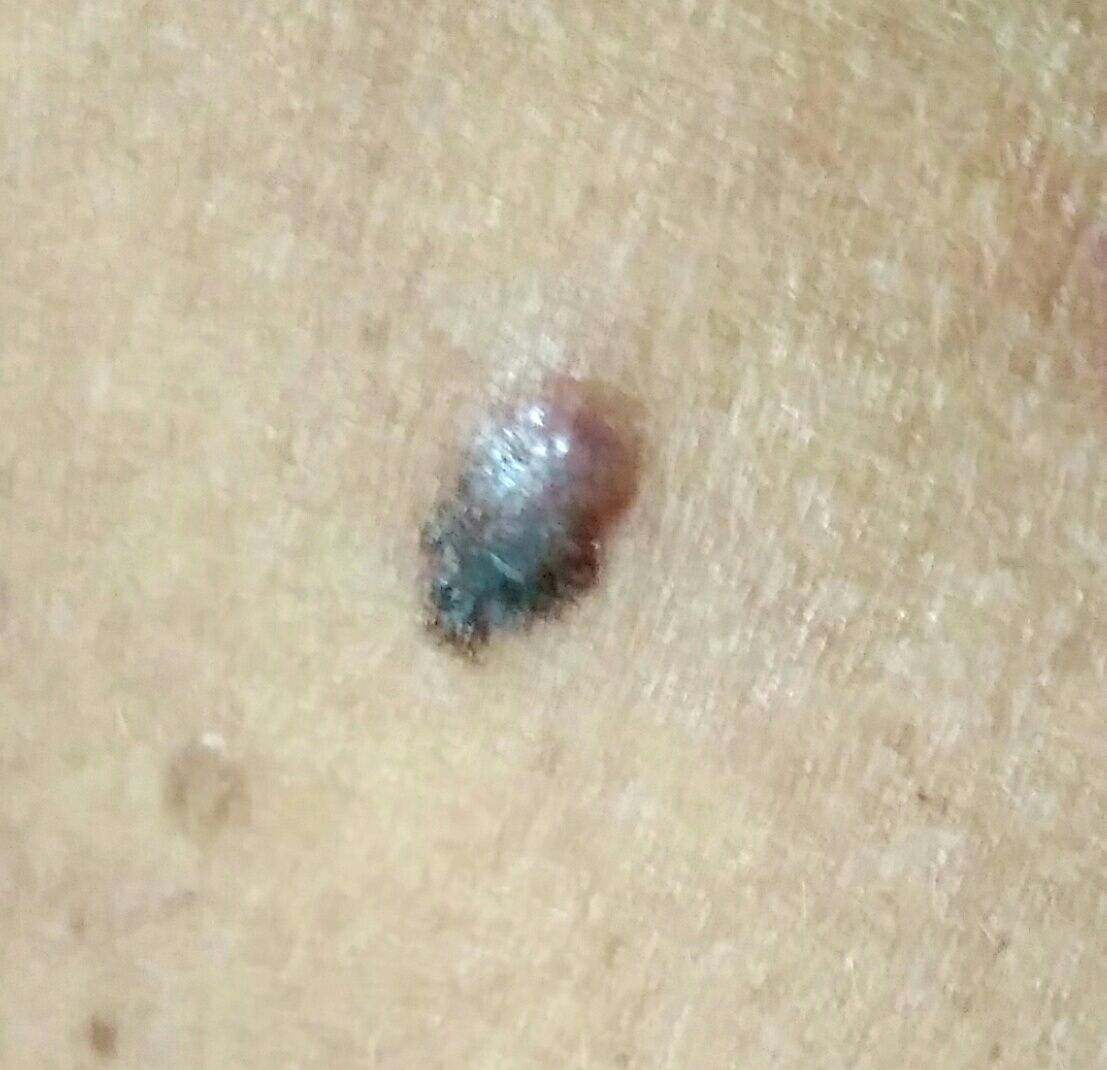
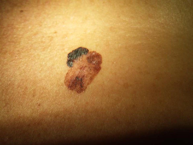
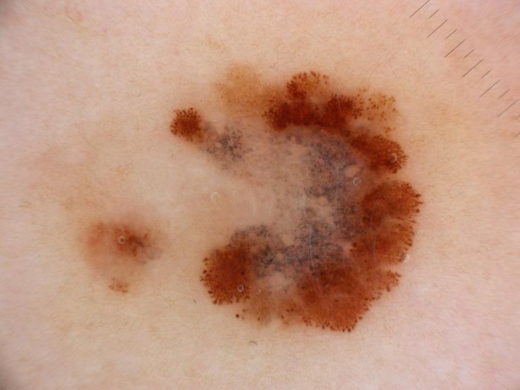
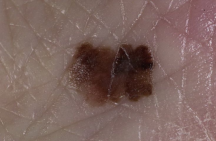
The color of melanoma is quite variable within the formation. The simultaneous presence of various shades of brown is possible: from light brown to dark, up to black. Dark, black shades usually prevail. The distribution of the pigment is heterogeneous (color heterogeneity over the entire area), asymmetric, with areas up to the complete absence of pigment (areas of regression, unfavorable factor). In addition to brown, other colors or their shades (polychrome) may be present: blue, blue, pink-red, white.
In the presence of other signs of malignancy, special alertness should be manifested in relation to formations over 5-6 mm. Or vice versa, any pigmented nevi with a diameter of more than 5 mm should be fully examined using the ABCDE system or dermatoscopically as potentially melanoma-hazardous formations. Most often, patients seek medical help with 8-15 mm melanomas, although there are tumors up to 10 cm.
The behavior of melanoma over time is rather mixed. Most often, this is a rapid growth in a few months. However, the horizontal growth phase (surface-spreading form) can last several years. Acceleration is observed during the transition to the phase of vertical growth. An assessment of the growth rate is quite informative when compared with the growth of other pigmented lesions (nevi) on the body. The evolution (change) of pigment formation also includes:
- The loss of previously present hair in the area of the age spots;
- The appearance of subjective sensations (itching, burning, tingling);
- Increase in tumor density;
- Surface changes (smoothing of the skin pattern, the appearance of ulcerations, cracks or exophytic component);
- Redness around the pigmented neoplasm;
- The rapid disappearance of part or all of the nevus, especially after ultraviolet radiation.
Later and neglected melanomas are characterized by:
- The appearance of nearby similar, but smaller foci (intradermal metastases);
- The presence of enlarged and dense lymph nodes along the lymphatic vessels (regional metastasis zone).
The presence of at least one of the above mentioneв symptoms is already an indication for consultation with a dermatologist or oncologist. The presence of three signs at the same time – the probability of melanoma reaches 80% or more.
By location on the body, melanomas are not selective. In women, the tumor is more often located on the legs, in men – on the trunk. In the elderly, melanomas of the skin of the face prevail.
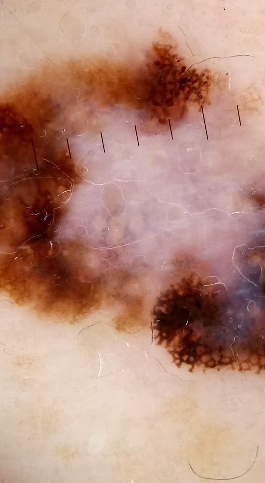
Dermatoscopic Description
With dermatoscopy of melanoma, multicomponentity is visualized (the simultaneous presence of a large number of different pathological patterns):
- Pigment network heterogeneity (atypical pigment network) – different intensity and eccentricity;
- Irregular stripes (mainly club-shaped);
- The presence of irregular inclusions of points and globules against the background of the pigment network (eccentric clusters);
- Globules of various shapes, sizes and colors;
- Asymmetry in color, structure and shape;
- Uneven edges;
- Peripheral radial radiance;
- Polychrome (3 colors);
- The presence of hypopigmentation zones and structureless zones, regression structures (unfavorable factor);
- Blue and white veil;
- Pathological vascular pattern (adverse factor).
Differential diagnosis
Differential diagnosis is carried out with such neoplasms as:
- Congenital dermal melanocytosis;
- Pigmented nevus (simple or papillomatous);
- Hemangioma (especially with vascular thrombosis);
- Blue nevus;
- Spitz Nevus;
- Dysplastic nevus;
- Lentigo;
- Pigmented basal cell carcinoma.
Risks
Melanoma is one of the most dangerous malignant tumors. In the world, approximately every 7 years, there is a double increase in the number of primary cases of melanoma. This is due, first of all, to an increase in the intensity of insolation and to a more frequent appearance of people in climatic zones unusual for their skin.
About 50% of all melanomas appear on healthy skin, the other half – against the background of existing pigmented benign neoplasms, which to some extent complicates the timely differential diagnosis and detection of malignant transformation.
Despite the fact that melanoma is approximately 10 times less common than other malignant skin tumors, mortality in the first case is 3.5 times higher.
Tactics
If you suspect or detect the first signs of melanoma, you need to consult an oncologist. The oncologist conducts additional specifying tests. In the absence of sufficient clinical data for an unambiguous diagnosis, sometimes the tactics of active dynamic observation is chosen. Most often, excision of a suspicious lesion is performed, followed by histological examination.
When confirming melanoma (clinically or histologically), a standard list of examinations is assigned to search for or exclude the presence of metastases, after which a special treatment plan is formed.
Treatment
In most cases, the treatment is surgical. Standard practice is to have a wide excision of melanoma under anesthesia or conduction anesthesia. If metastases are detected in regional lymph nodes, lymph dissection is performed (removal of the entire block of regional lymph nodes). If distant metastases are detected, the treatment regimen is selected individually. For this, the oncologists’ arsenal has quite effective chemotherapy, immunotherapy, radiation therapy regimens, as well as the possibility of surgical removal or minimally invasive therapy of metastases.
Treatment of melanoma (even the earliest forms) with local destruction methods (laser removal or cryodestruction) or removal under local anesthesia is unacceptable.
Prevention
Prevention of the appearance of melanoma is a gentle and careful attitude to the skin:
- Limitation of ultraviolet radiation (naturan (sun) and artifitial);
- The use of protective creams during periods of active sun;
- Exclusion of chronic skin trauma;
- Limitation or exclusion of ionizing radiation, occupational hazards;
- Compliance with safety measures when working with skin damaging factors;
- Personal hygiene and basic awareness of skin tumors.
It also requires regular examination of all pigmented neoplasms, timely consultation of a specialist in the event of external changes, and removal of potentially dangerous skin tumors.