Dermatofibroma – (skin fibroma, benign fibrous histiocytoma) benign neoplasm of the connective tissue genesis (non-melanocytic), represented by a knot, towering above the skin or intradermal dense knot. Dermatofibroma is an acquired neoplasm, it is extremely rare in newborns and children, as a rule – they appear in people older than 25-30 years. The multiplicity of the lesion is not characteristic (with the exception of some systemic diseases, hereditary syndromes, and HIV infection). By gender: dermatofibromas are more common in women.
Predisposing factors
There is no clear reason for the appearance of dermatofibromas. It is only appropriate to talk about predisposing factors that, to varying degrees, can increase the risk of neoplasms:
- Female gender;
- Damage to the skin, microtrauma, insect bites;
- Genetic factor: heredity can play a role in the appearance of dermatofibromas.
Diagnostics
The diagnosis of dermatofibroma is based on a clinical examination, which includes a routine examination of the formation and dermatoscopy. If you suspect malignant growth, a biopsy can be performed.
Symptoms
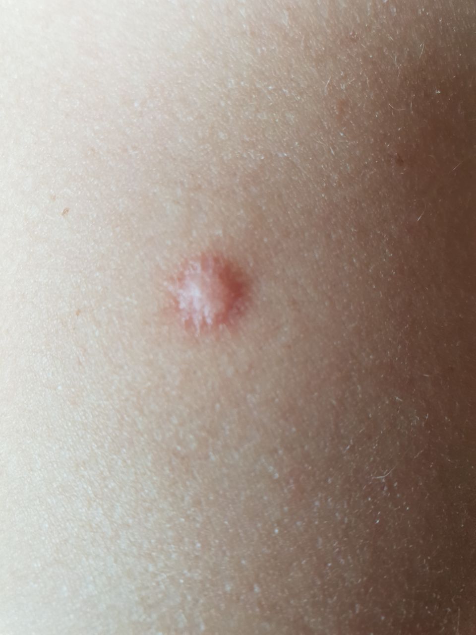
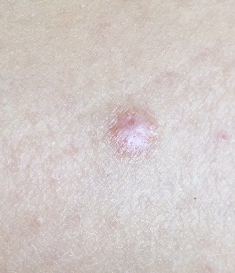
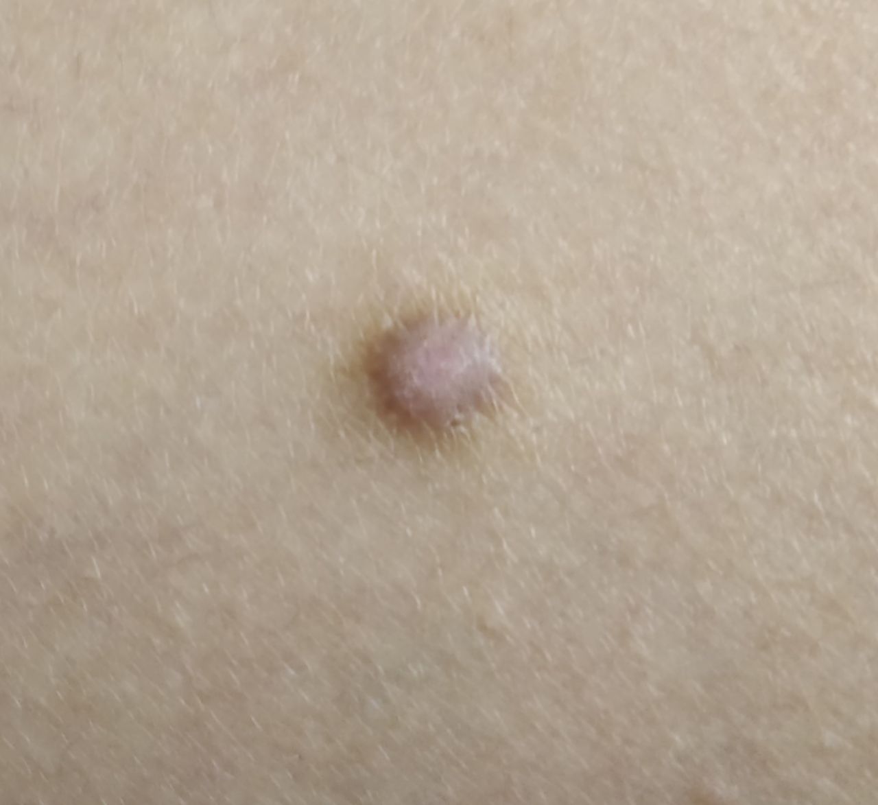
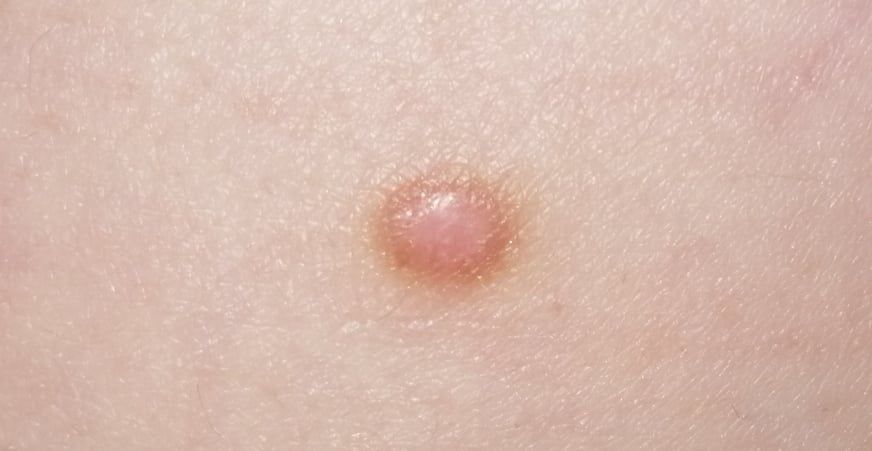
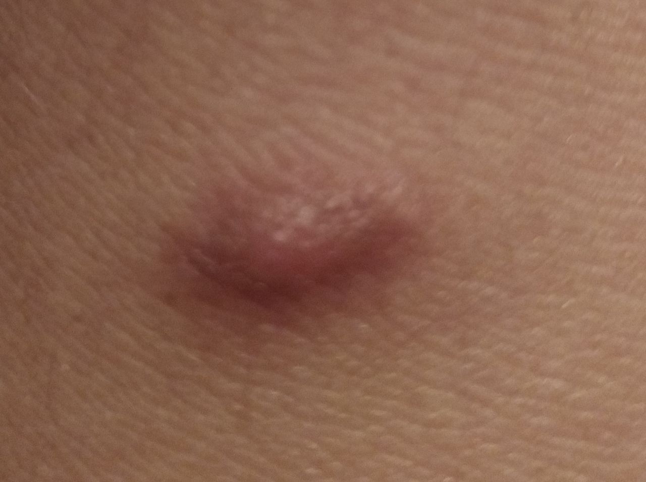
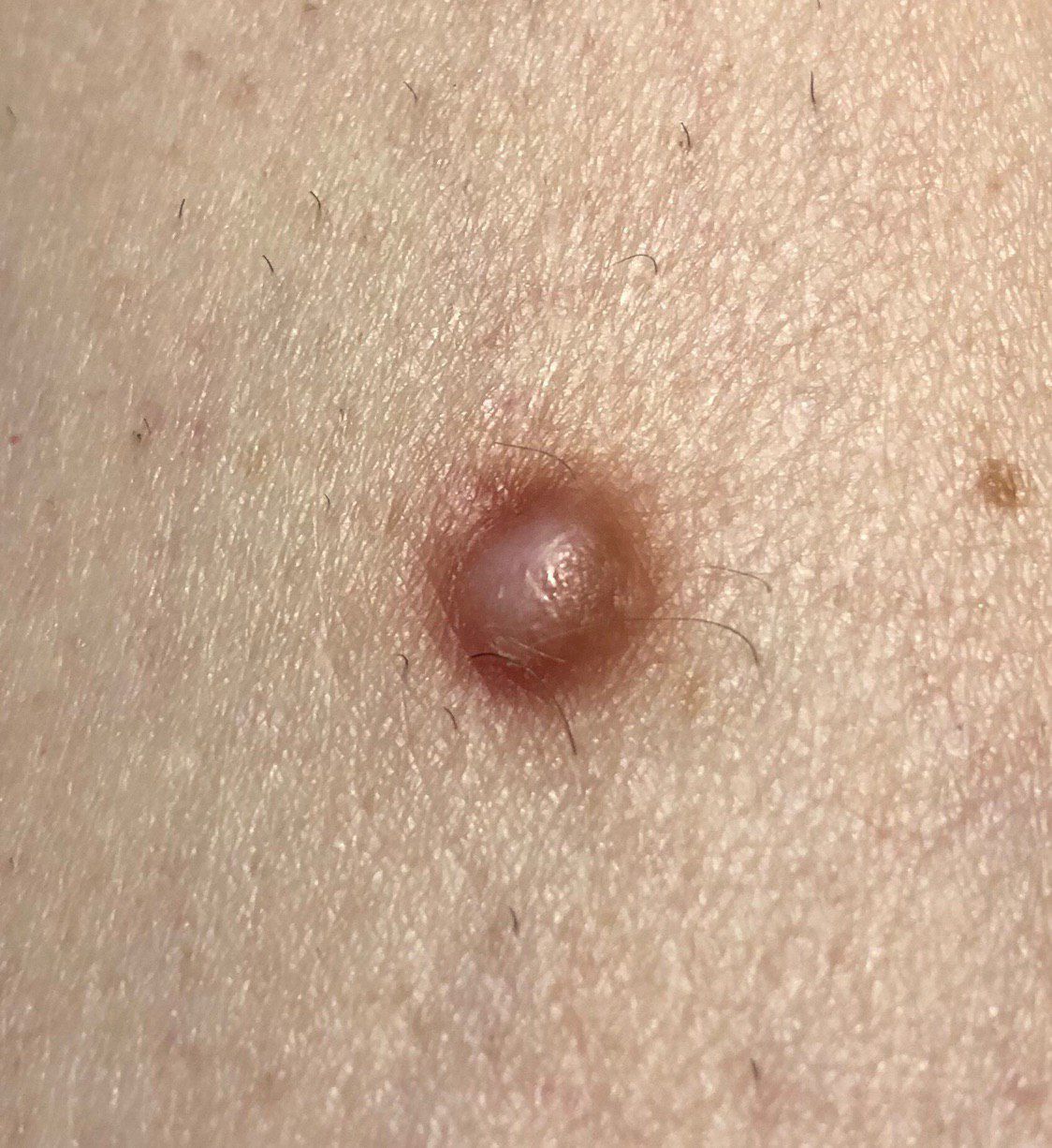
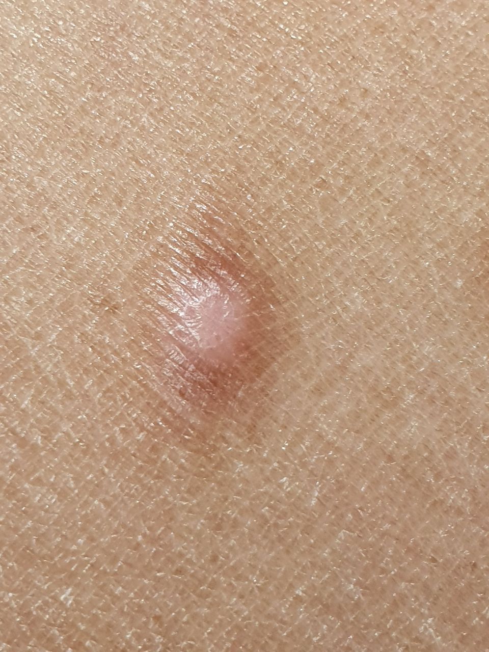
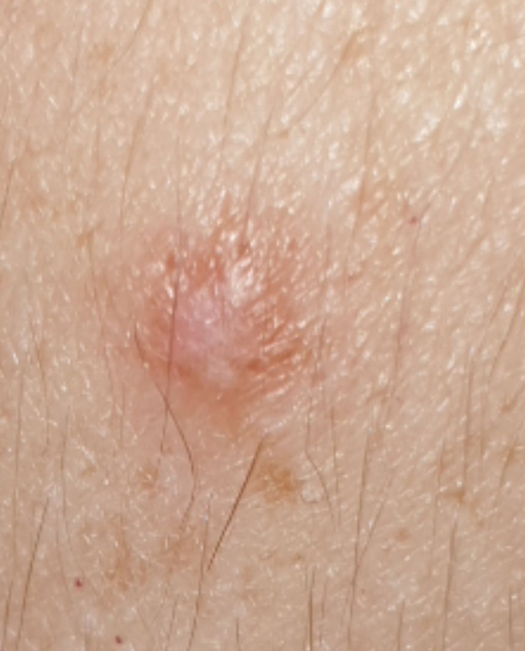
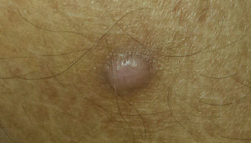
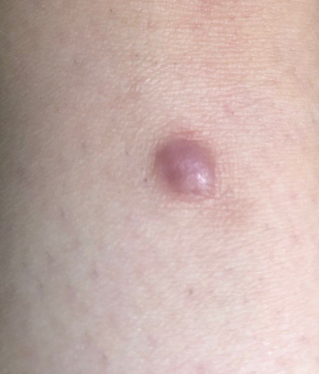
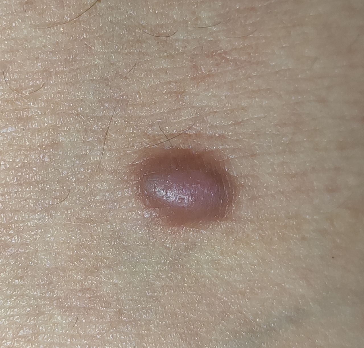
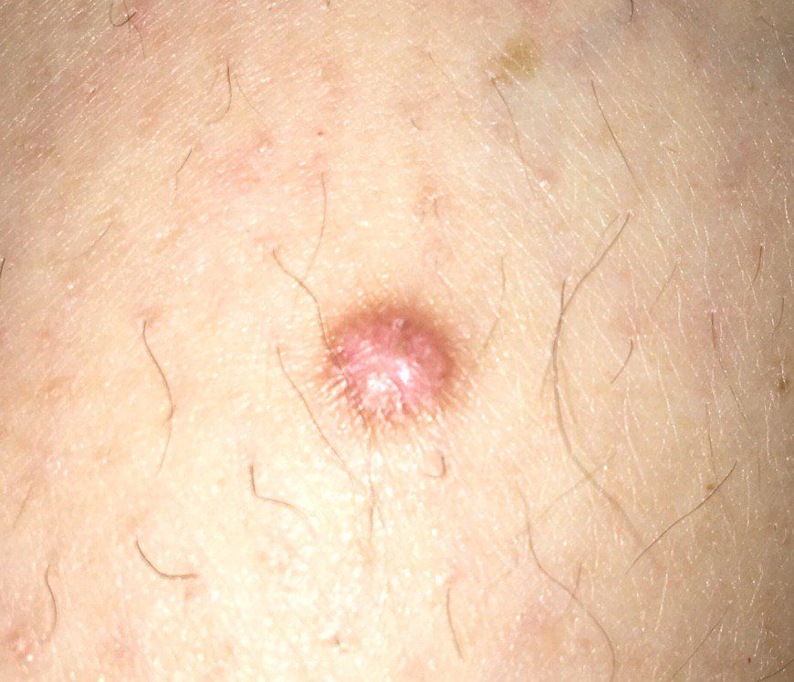
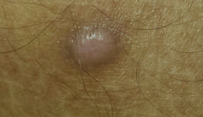
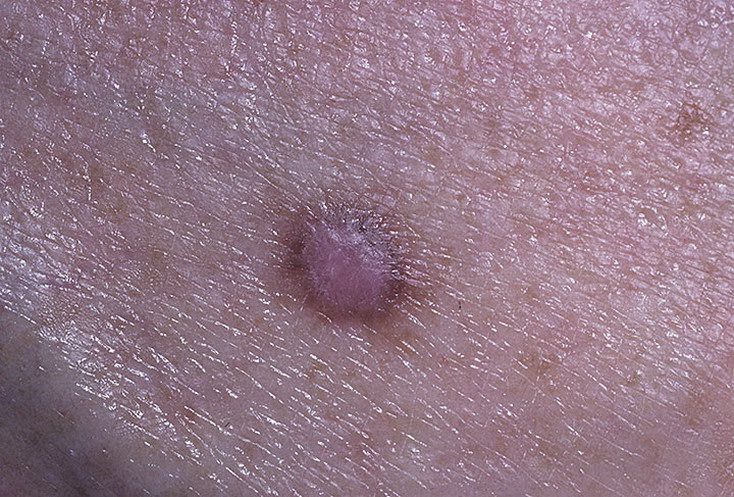
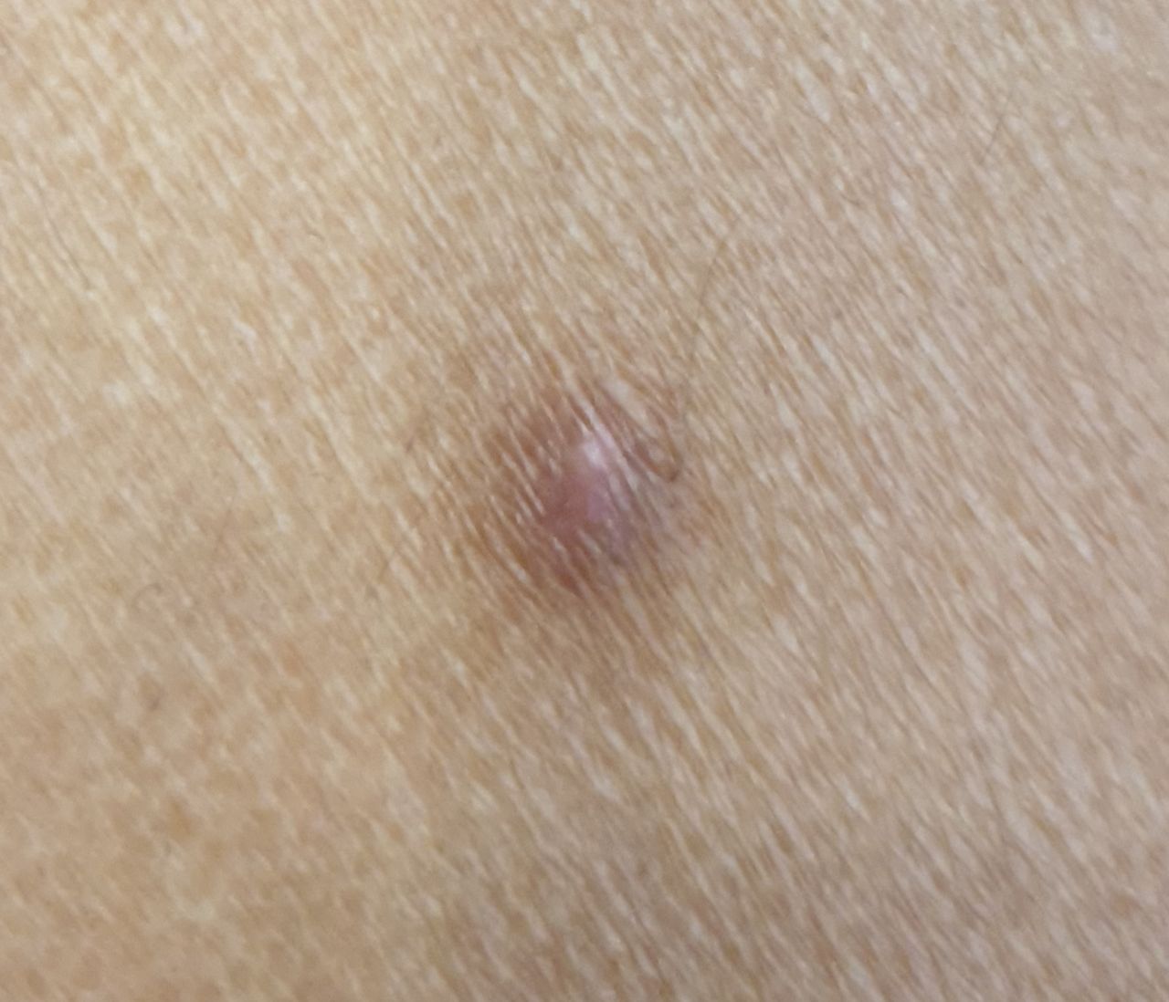
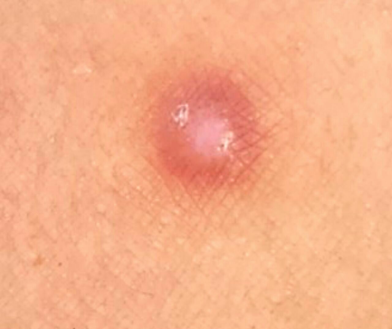
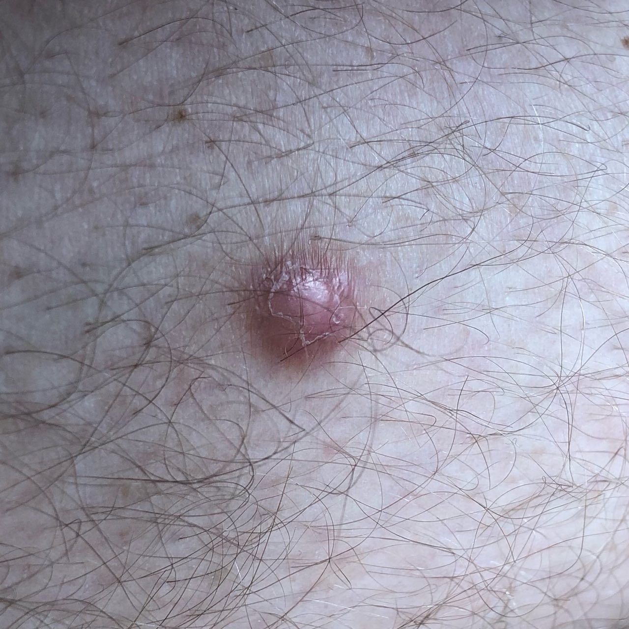
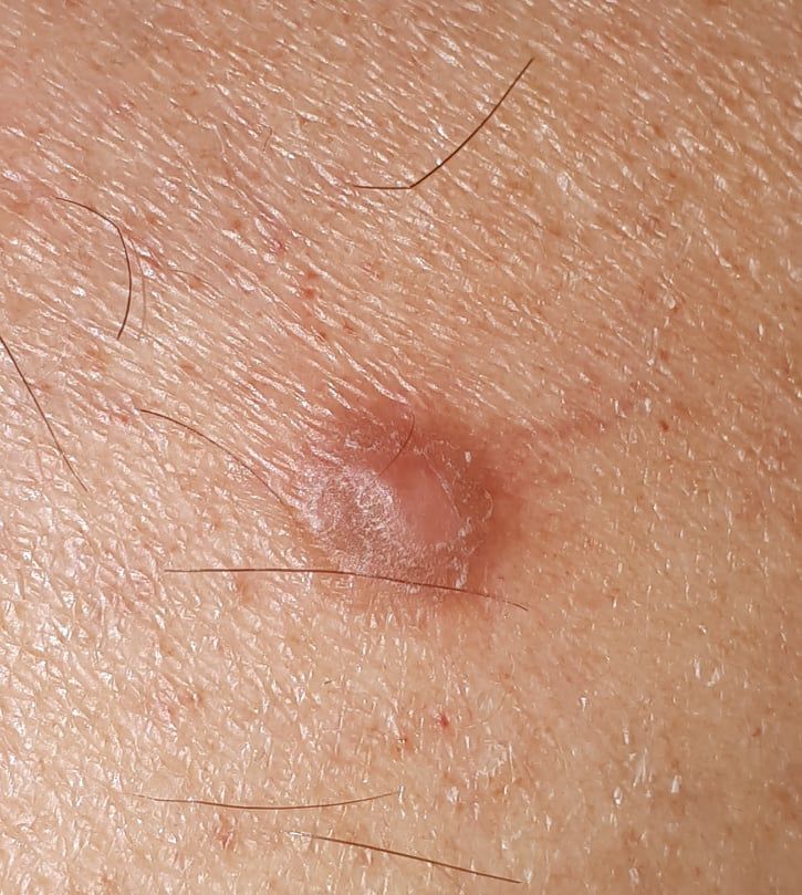
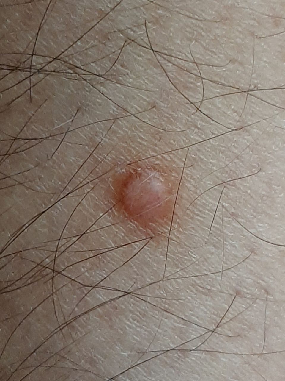
A visual examination of the dermatofibroma determines a slightly sunken (below the surface of the skin) or hemispherical formation, most often symmetrical (oval or round). The surface of the dermatofibroma is usually smoothed, the skin pattern is most often absent (especially in the center), less often small tuberosity, peeling.
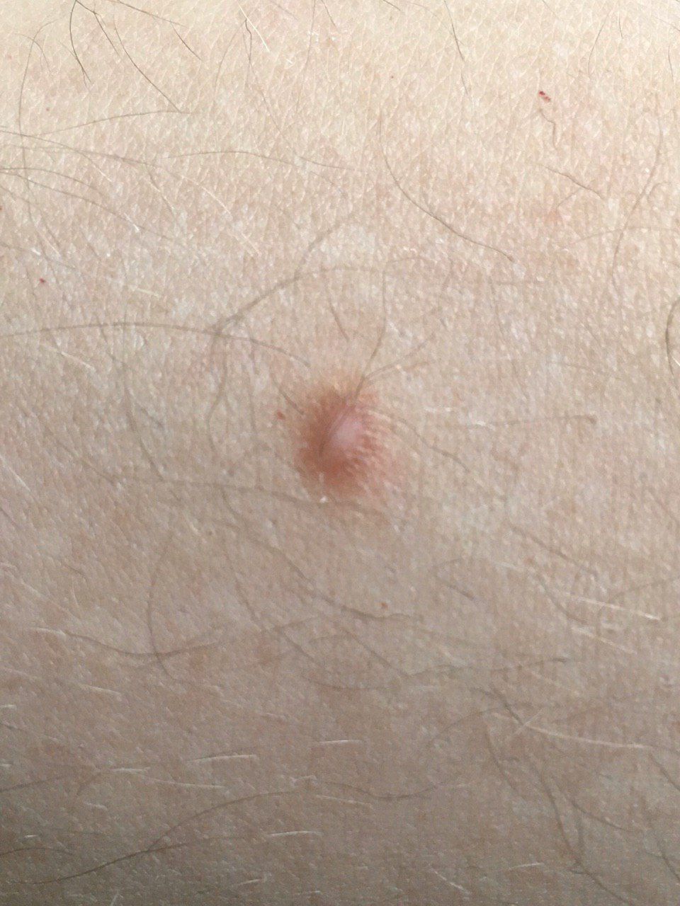
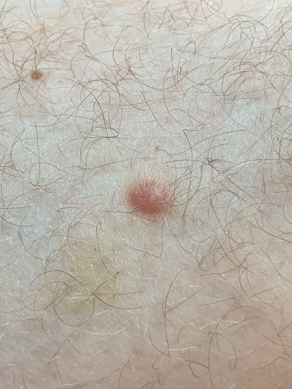
The boundaries of the dermatofibroma can be clear and even (in protruding hemispherical forms) or blurry (if the neoplasm is flat or sunken). Color varies from flesh, gray to shades of brown and crimson. The distribution of pigment throughout the formation is uneven: a gradual increase in color intensity is observed in the direction from the center to the periphery (the center of formation is lighter, even pale, the periphery of the formation is more intensely colored).
Hair in the central part of dermatofibroma is absent, but can occur on the periphery.
The diameter of the dermatofibroma usually does not exceed 10 mm (larger formations are rarely found). The height above the skin for hemispherical forms is usually 5-7 mm.
On palpation, the dermatofibroma is dense, firm. A distinctive feature is a symptom of the fossa: when the surrounding tissues are compressed, a depression is formed in the tumor area. There is no subjective sensation or mild itching (rarely).
They are located mainly on the lower extremities and in the shoulder girdle, other localizations are rarely observed.
Dermatoscopic Description
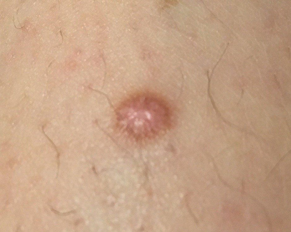
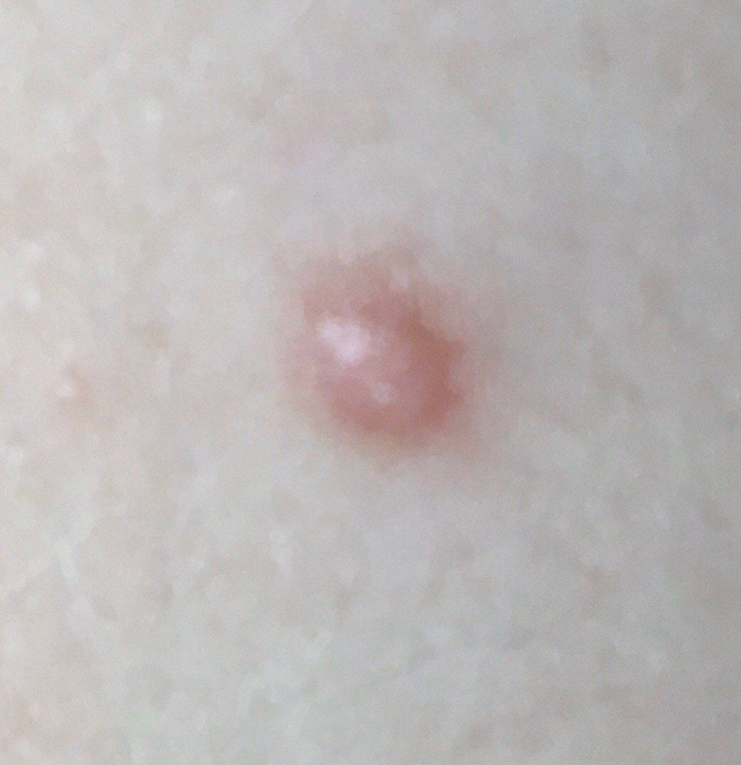
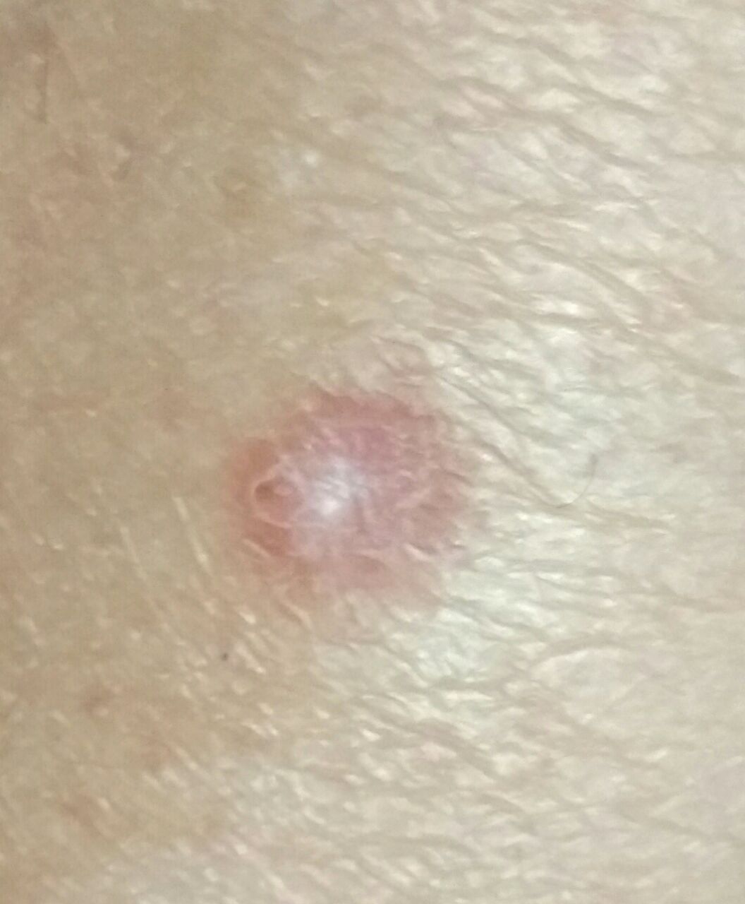
With dermatoscopy, the following dermatofibroma features are visualized:
- Hypopigmentation in the central zone;
- Incorrect contour of the hypopigmented central zone;
- A light brown peripheral zone that surrounds the central zone;
- The peripheral zone is represented by a delicate pigment network with small cells;
- The boundaries of the peripheral zone smoothly pass into healthy skin;
- Fine-meshed uniform structure at the border of transition to healthy skin;
- Point vessels may rarely be detected.
Differential diagnosis
Differential diagnosis is carried out with such neoplasms as:
- Simple nevus
- Papillomatous nevus
- Comedones
- Hemangioma
- Blue nevus
- Dysplastic nevus
- Keratoacanthoma
- Basal cell carcinoma
- Nodular melanoma
- Swelling dermatofibrosarcoma
Risks
Dermatofibroma is safe and does not carry an increased risk of a malignant tumor. In the absence of external influences (trauma and other damaging factors) – the risk of malignant degeneration is comparable to the same risk on unchanged skin.
Signs of a possible malignancy: a change in appearance, the appearance of subjective sensations.
Tactics
In the absence of any damaging effect on the dermatofibroma, changes in appearance and subjective sensations, removal is not necessary, self-control (or examination with the help of other persons in inaccessible areas) is sufficient at least once a year. If there was a mechanical damage to the dermatofibroma, changes in appearance were noticed, or previously absent sensations appeared, a dermatologist or oncologist should be consulted.
The specialist determines the possibility of further dynamic monitoring (terms are determined individually) or indications for removal are set. It is necessary to remove those dermatofibromas that are subject to constant, chronic trauma to clothing, jewelry, or due to the characteristics of professional employment.
In the case of dynamic observation, photo fixation of skin neoplasms is of great value, which will subsequently determine even minor changes in appearance.
Patients, in the presence of other skin neoplasms (nevi) in combination with dermatofibroma, are shown an examination by a dermatologist or oncologist in the spring and autumn (before and after the beach season). Such patients are recommended to compile a map of skin neoplasms, which greatly simplifies further observation, the search for new formations, or a change in existing ones.
Treatment
Only surgical (classic, using an electric or radio scalpel) with a mandatory histological examination.
Dermatofibroma treatment using destructive methods (laser removal or cryodestruction) is not recommended, since in this case there is a pronounced tendency to local relapses.
Prevention
Prevention of the appearance of dermatofibromas and their malignancy consists in a gentle and careful attitude to the skin:
- Exclusion of chronic skin trauma;
- Compliance with safety measures when working with skin-damaging factors;
- Personal hygiene and basic awareness of skin tumors.
Also, a regular examination of dermatofibromas, timely consultation of a specialist in the event of external changes, and the removal of potentially dangerous neoplasms are necessary.