Simple nevus (benign nevus, pigmented nevus, mole, birthmark) – a benign neoplasm of the skin, which is a spot or a small nodule that rises slightly above the skin. Simple nevus can be either congenital or acquired (a characteristic appearance at any age). Approximately 3% of newborns have a plurality of simple nevi. With age, the proportion of multiplicity increases. By gender, simple nevi are somewhat more common in women compared with men, in a ratio of 3: 2, respectively.
Predisposing factors
There is no clear reason for the appearance of simple nevi. It is only appropriate to talk about predisposing factors that, to varying degrees, can increase the risk of neoplasms:
- Genetic factor: the appearance of pigmented nevus may be due to the human genome;
- Ultraviolet radiation: artificial or solar ultraviolet leads to faster reproduction of nevoid cells (nevus cells) and excessive production of melanin (pigment, the accumulation of which is noted in nevus);
- Hormonal changes: hormonal fluctuations in the body (especially sex hormones, thyroid hormones and adrenal hormones) can affect the appearance of new nevi and the growth of existing ones;
- Ionizing radiation, viral diseases and injuries can also trigger the appearance or growth of simple nevi.
Diagnostics
Diagnosis of simple nevi is based on a clinical examination, which includes a routine examination of the formation and dermatoscopy. If you suspect malignant growth, a biopsy can be performed.
Symptoms
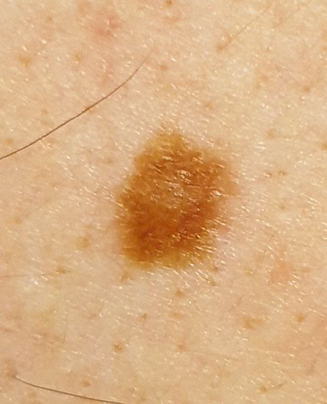
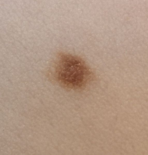
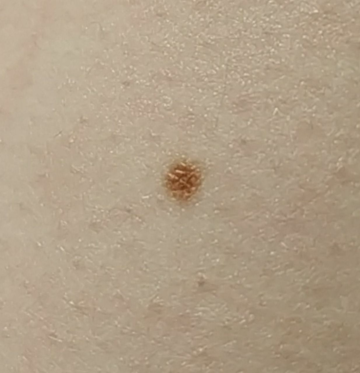
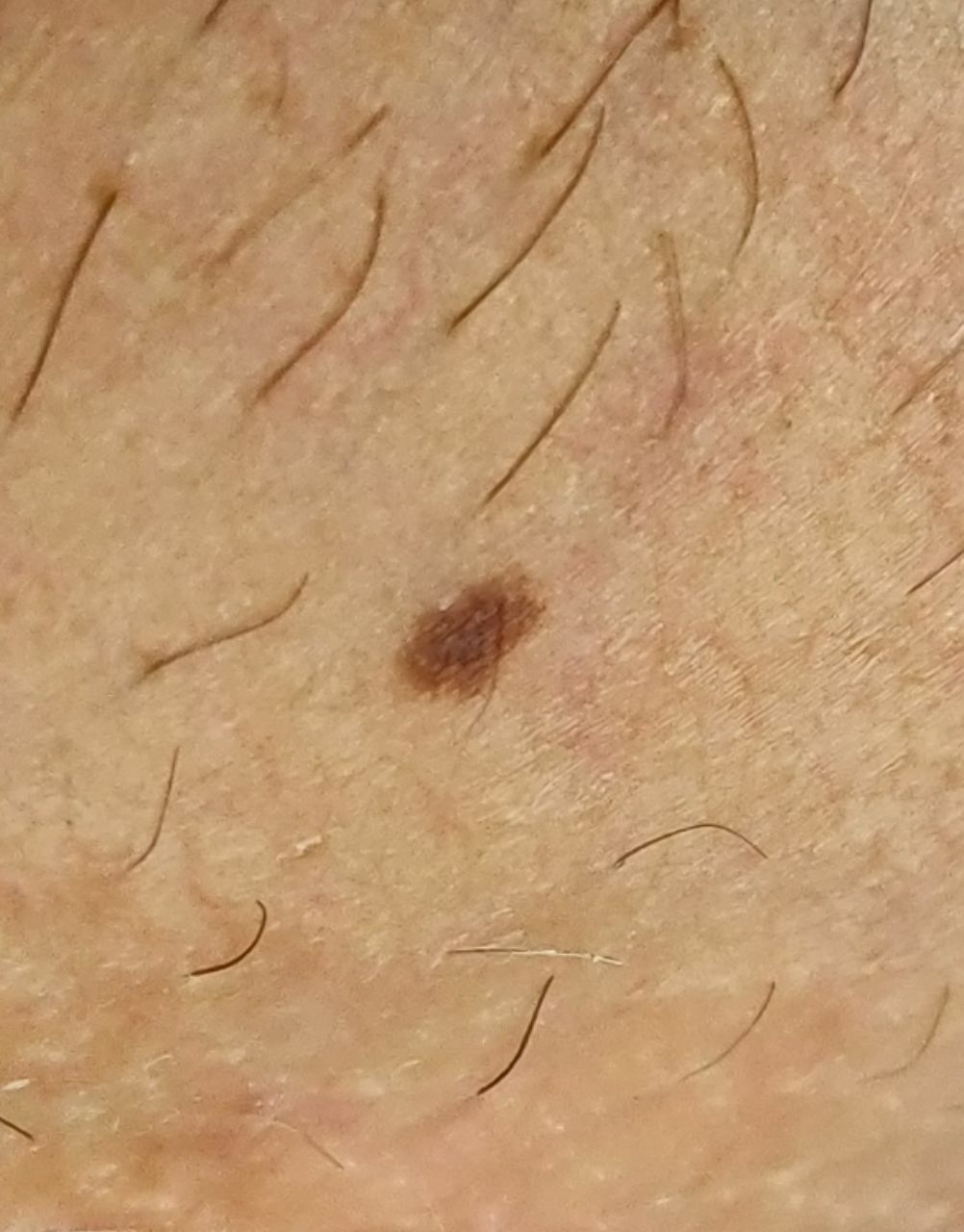
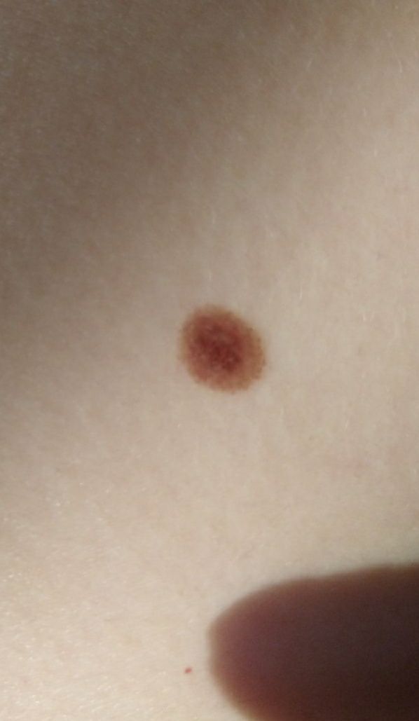
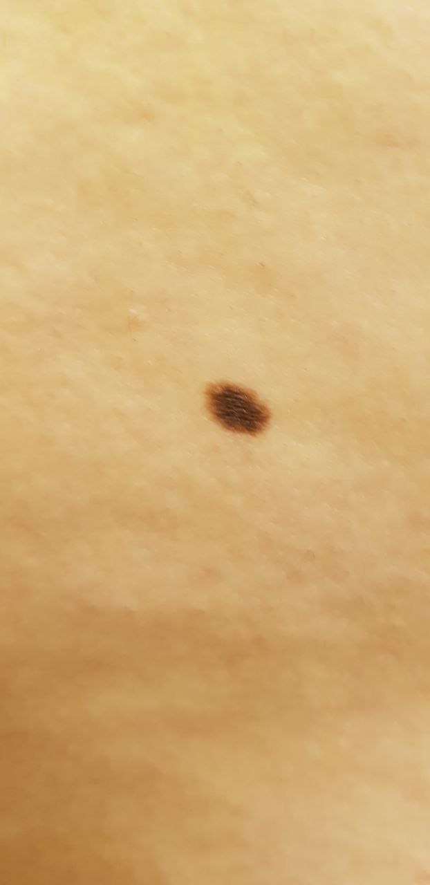
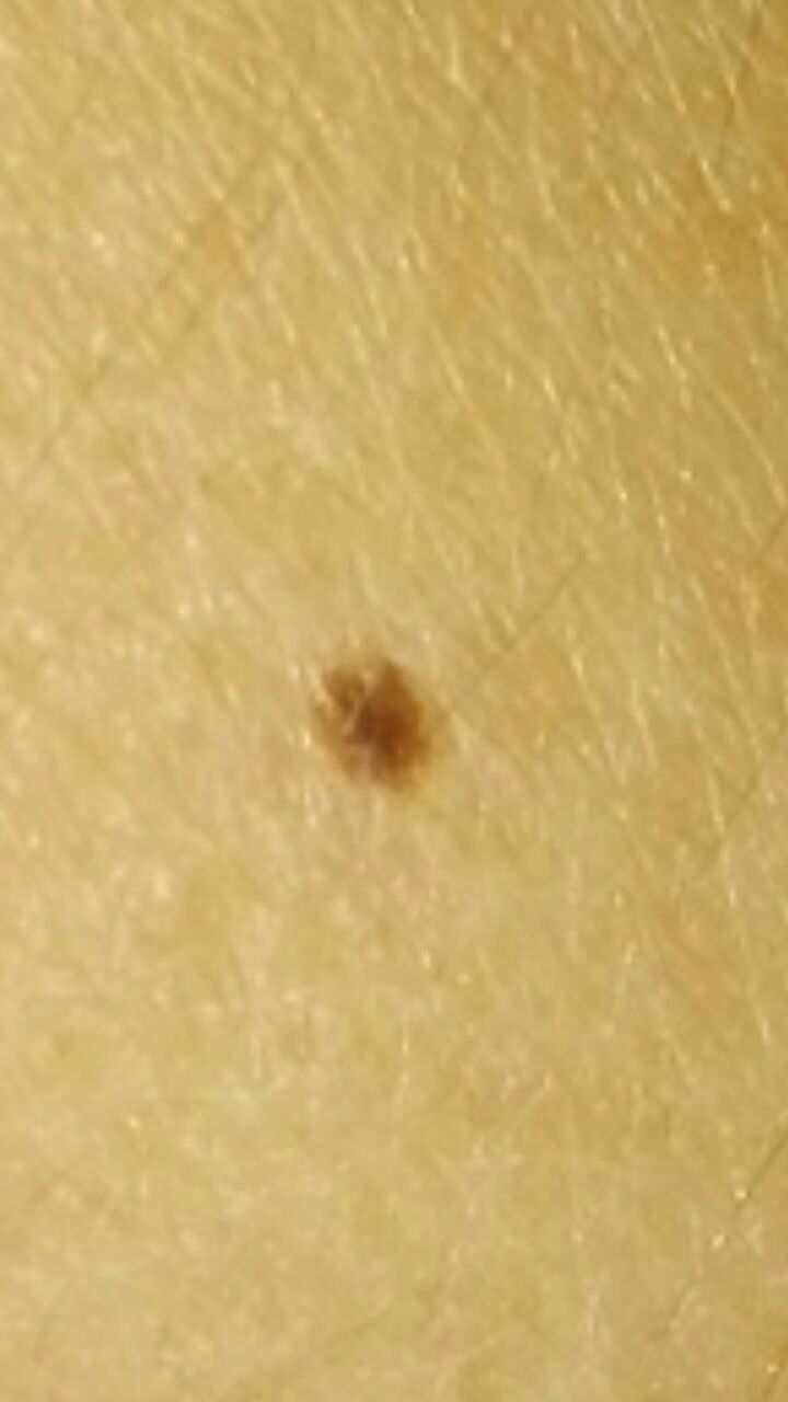
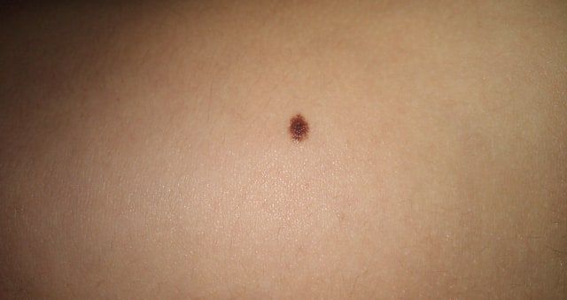
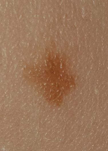
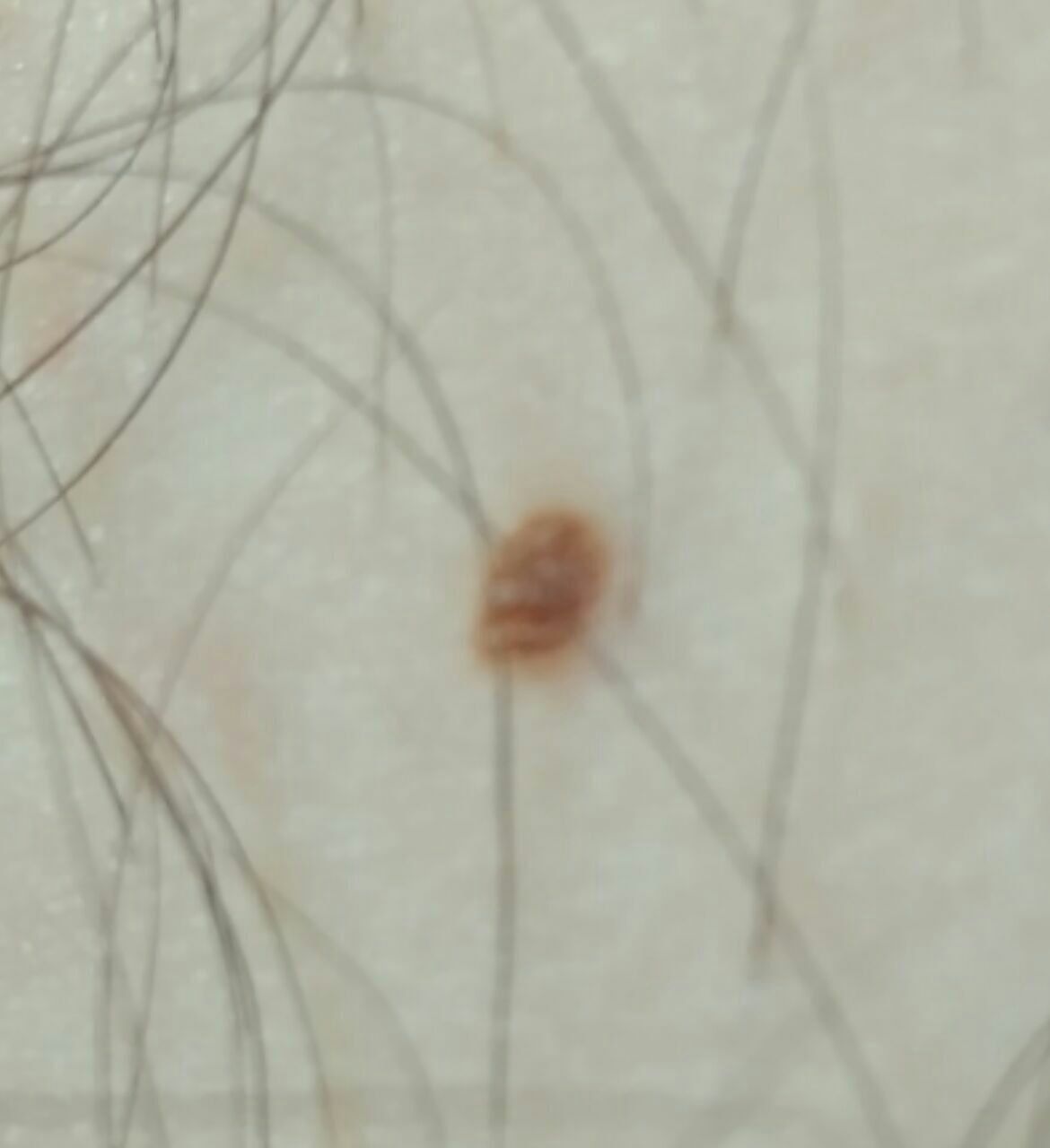
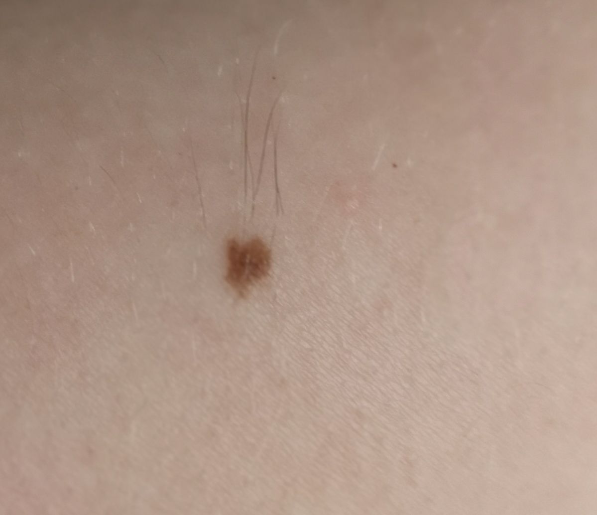
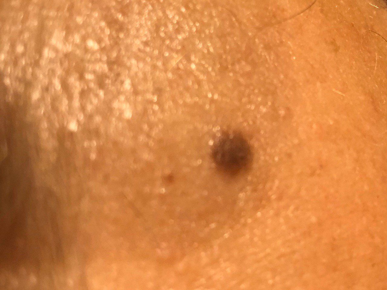
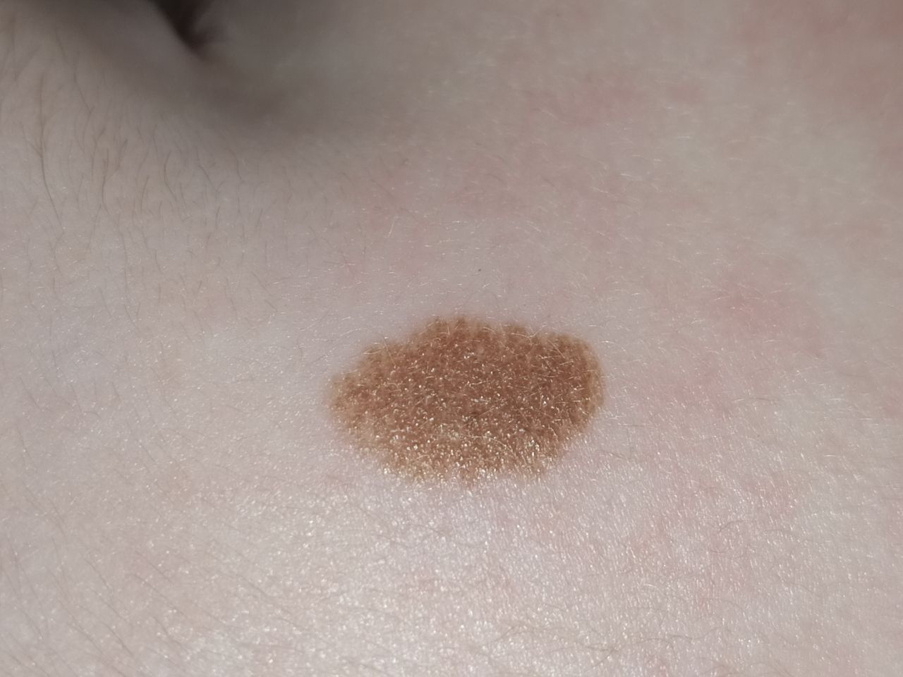
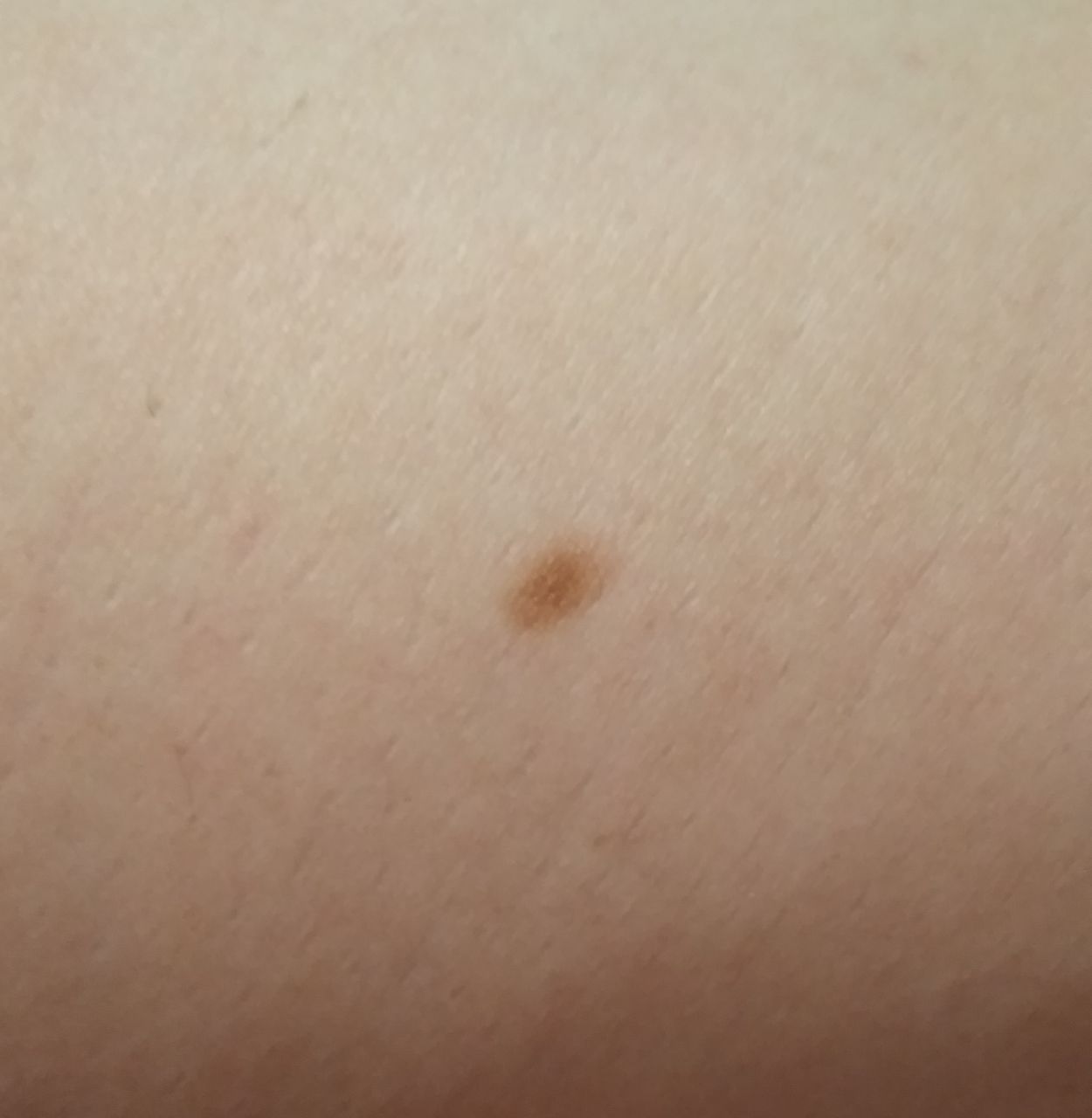
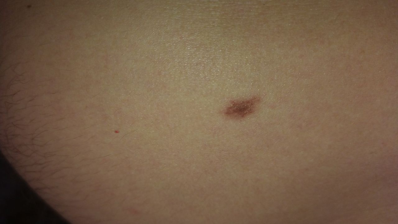
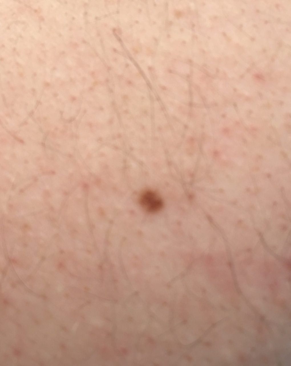
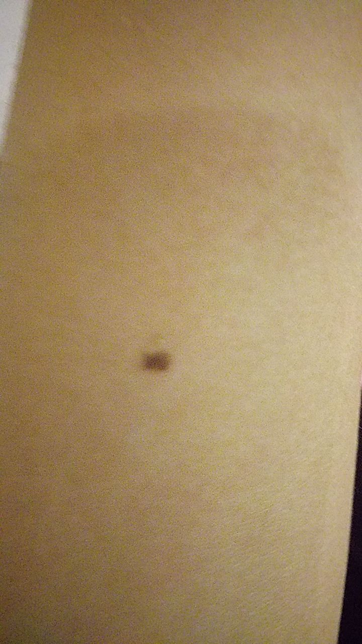
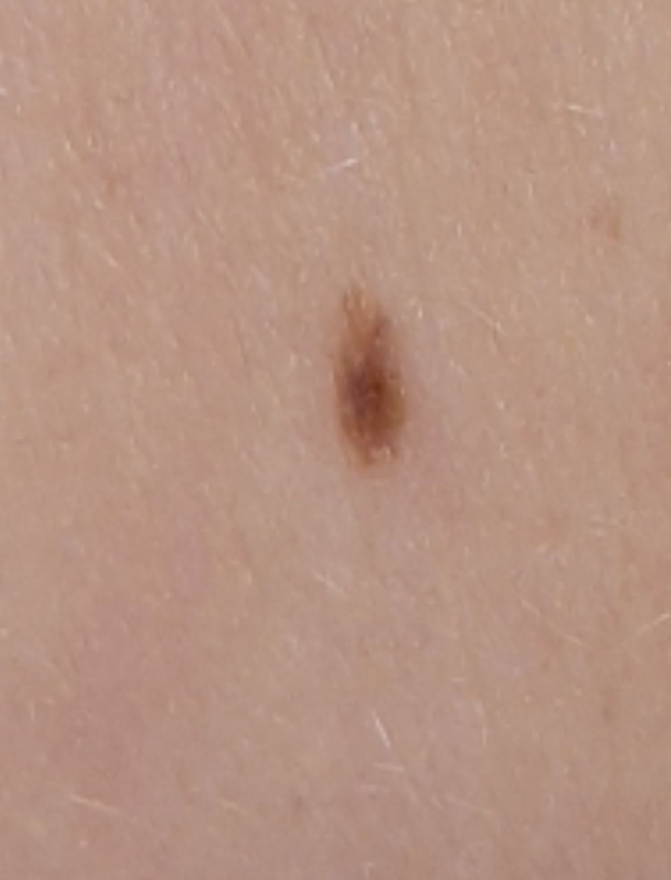
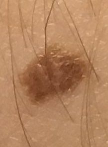
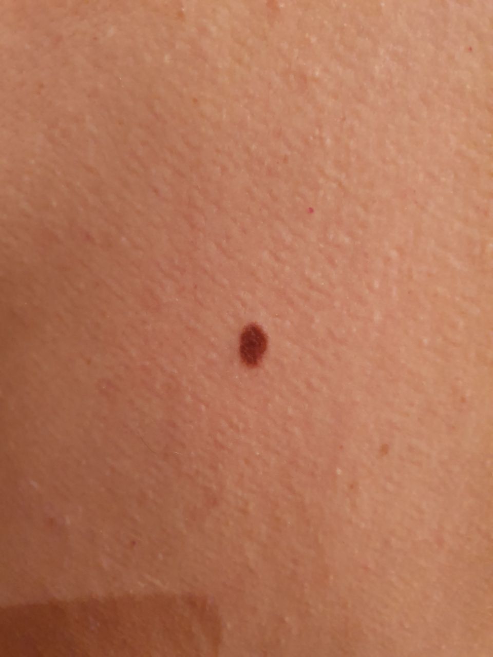
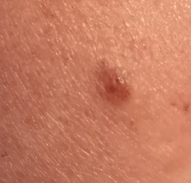
On visual inspection of a simple nevus, a spot or slightly elevated nodule is determined. Most often, the formation is symmetrical (oval or round), however, congenital nevi of large sizes can also be irregular in shape. The surface of the nevus is a texture of ordinary skin (rarely – slightly differs from the skin pattern).
The boundaries of benign nevus are clear and even. Large and giant congenital nevi can be with uneven edges. The color of simple nevus varies from tan to dark brown, with the distribution of pigment throughout the formation uniform. Sometimes there is a gradual decrease in color intensity in the direction from the center to the periphery. The shades of the congenital nevus can change in the first years of a person’s life.
The presence of benign nevus does not affect hair growth. Less often in the area of congenital nevus there is a more intensive growth of coarse bristly hair, which is usually combined with pronounced brown pigmentation.
The sizes of simple benign nevi can vary within wide limits, however, the most common formations are up to 10 mm. Nevi over 10 mm – in most cases congenital, they are rare, can reach 20 cm or more (giant congenital nevi).
On palpation of a simple nevus, there are no features: the consistency of ordinary skin. Subjective sensations are also absent.
Neoplasms are located mainly on the trunk (~ 38%) or limbs (~ 48%), less often on the head and neck (~ 14%).
Nevi of the plantar and palmar surfaces (acral nevi) are somewhat different in shape, contour, and pigment distribution from ordinary nevi, which is associated with the presence of a characteristic skin pattern (“fingerprints”). Nevi of this localization are elongated in shape, have an uneven contour, darker color and pigment distribution in the form of parallel stripes.
Dermatoscopic Description
With dermatoscopy of a simple nevus, the following features are visualized:
- The pigment network is a pattern of hypopigmented holes and homogeneous lines from light brown to dark brown. The lines are evenly thinning to the periphery of the formation;
- Dots – are small hyperpigmented round structures that are located in the center or are found on the pigmented lines of the network;
- Globules – large hyperpigmented ring structures that are evenly distributed throughout the nevus or in the center, are rarely found on the periphery;
- Spots – hyperpigmented structureless areas located in the center;
- Vascular network – represented by slightly curved diffuse monomorphic vessels;
- Star radiance – pigmented stripes and dots located in the form of rays on the periphery;
- Diffuse uniform staining of the entire formation
Acral nevi have some features in dermatoscopic imaging:
- Accumulation of nevus cells is mainly found in furrows, and atypical melanocytes in ridges. The eccrine duct opens to the epidermis through ridges, which can help distinguish the ridge from the groove;
- The main pattern is trellised, parallel, and fibrillar. The fibrillar pattern is more common with the plantar surface of the foot (at the site of the greatest application of body weight).
Differential diagnosis
Differential diagnosis is carried out with pigmented neoplasms such as:
- Post-inflammatory hyperpigmentation
- Congenital dermal melanocytosis
- Nevus of the sebaceous glands
- Halo nevus
- Spitz Nevus
- Blue nevus
- Lentigo
- Dysplastic nevus
- Lentigo melanoma
- Melanoma
Risks
Simple nevus is safe and does not carry an increased risk of melanoma. In the absence of external effects on the pigmented nevus (trauma, ultraviolet radiation, ionizing radiation) – the risk of malignant degeneration is comparable to the risk of melanoma on unchanged skin. Signs of a possible malignancy of a simple nevus: a change in appearance, the appearance of subjective sensations.
A slightly increased risk of melanoma is observed in congenital nevus, but this is more typical for large pigmented lesions (more than 20 cm in diameter). The risk of melanoma on the background of congenital nevus up to 20 cm is less than 1%.
Large and multiple congenital nevi can be associated with various genetic syndromes and diseases, therefore, such patients require more careful observation and examination.
Tactics
In the absence of any damaging effect on the simple nevus, changes in appearance and subjective sensations – self-control (or examination with the help of other persons in inaccessible areas) is enough at least once a year. If mechanical damage to the nevus has occurred, its active irradiation with ultraviolet or ionizing radiation, as well as if any changes in the nevus itself are noticed or previously absent sensations appear, you need to consult a dermatologist or oncologist.
The specialist determines the possibility of further dynamic monitoring (terms are determined individually) or indications are made for removal of the damaged nevus. It is necessary to remove those nevi that are subject to constant, chronic trauma to clothing, jewelry, or due to the characteristics of professional employment.
In the case of dynamic observation, photo fixation of skin neoplasms is of great value, which will subsequently determine even minor changes in the appearance of the nevus.
Patients with congenital or multiple acquired moles require examination by a dermatologist or oncologist in the spring and autumn (before and after the beach season). Such patients are also recommended to compile a map of skin neoplasms, which greatly simplifies further observation, the search for new formations, or a change in existing ones.
Treatment
Only surgical (classic, using an electric or radio scalpel) with a mandatory histological examination.
Treatment of pigmented nevus using destructive methods (laser removal or cryodestruction) is not recommended.
Prevention
Prevention of the appearance of nevi and their malignancy consists in a gentle and careful attitude to the skin:
- Limitation of ultraviolet radiation (tanning bed, solar tanning);
- The use of protective creams during periods of active sun;
- Exclusion of chronic skin trauma;
- Limitation or exclusion of ionizing radiation, occupational hazards;
- Compliance with safety measures when working with skin-damaging factors;
- Personal hygiene and basic awareness of skin tumors.
Also, a regular examination of the pigmented nevus, timely consultation of a specialist in the event of external changes, and removal of potentially dangerous neoplasms are necessary.