Pyogenic granuloma (telangiectatic granuloma, botriomycoma, granulation Tissue-type Hemangioma, lobular capillary hemangioma, eruptive angioma, inflammatory hemangioma) is a benign neoplasm, which is a local reaction of blood capillaries in response to an external effect (most often an injury). Pyogenic granulomas appear in various parts of the body, including the mucous membranes, conjunctiva, and cornea. It is more common among young people as well as pregnant women.
Predisposing factors
There is no clear reason for the appearance of pyogenic granulomas. Among etiological factors, there are many versions. Previously, injuries (point injuries, cuts, foreign bodies, burns) were considered the main cause. However, today it is established that only 25% of all pyogenic granulomas appear against the background of injuries. Other possible causes include:
- Infectious skin diseases;
- Dermatoses
- The presence of large burn surfaces;
- Taking oral contraceptives;
- Reception of protease inhibitors;
- Isotretinoin Acne Treatment;
- Pregnancy (Increased blood concentration of growth factors).
Diagnostics
Diagnosis of pyogenic granulomas is based on a clinical examination, which includes a routine examination of the formation and dermatoscopy. If you suspect malignant growth, a biopsy can be performed.
Symptoms
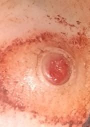
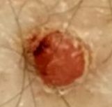
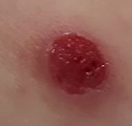
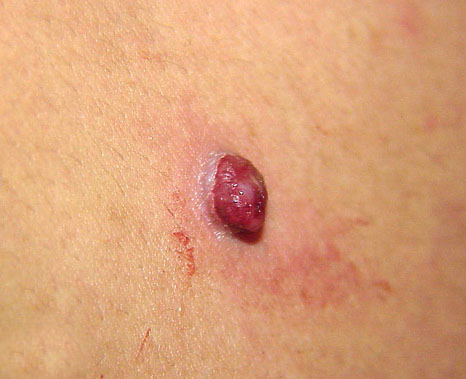
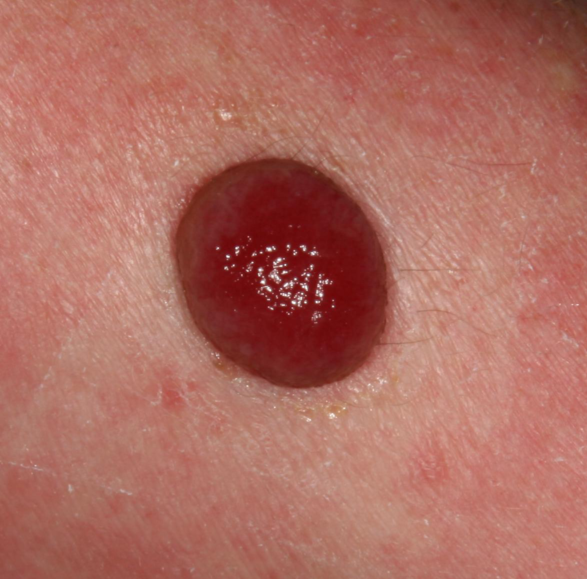
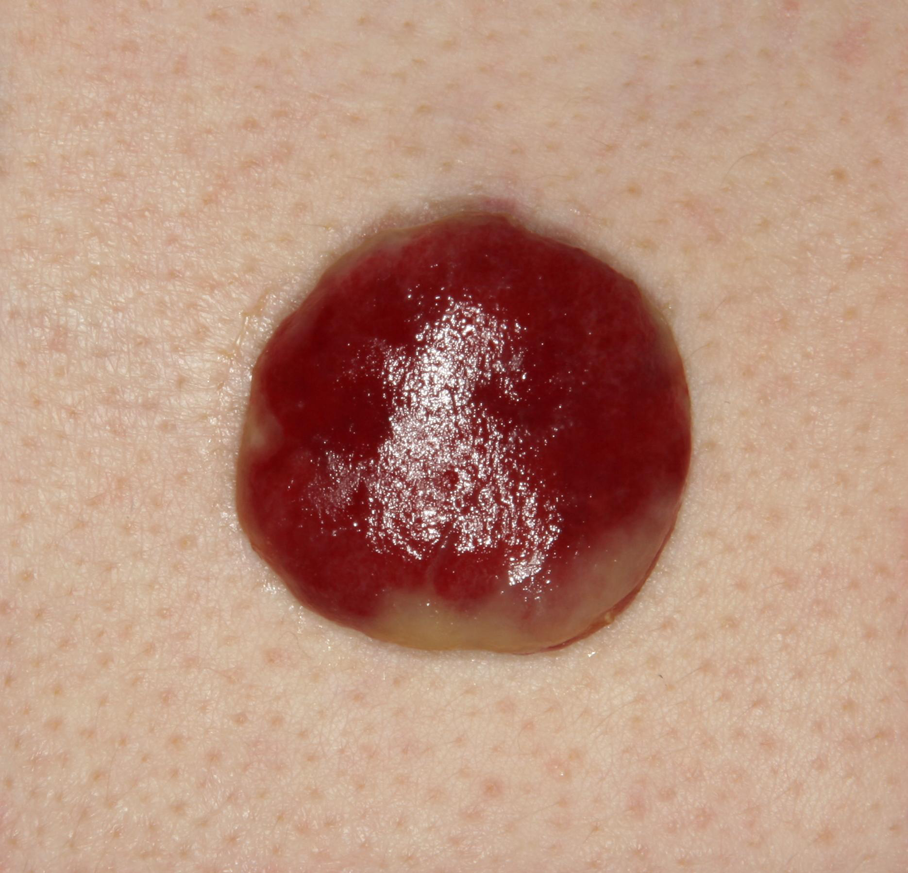
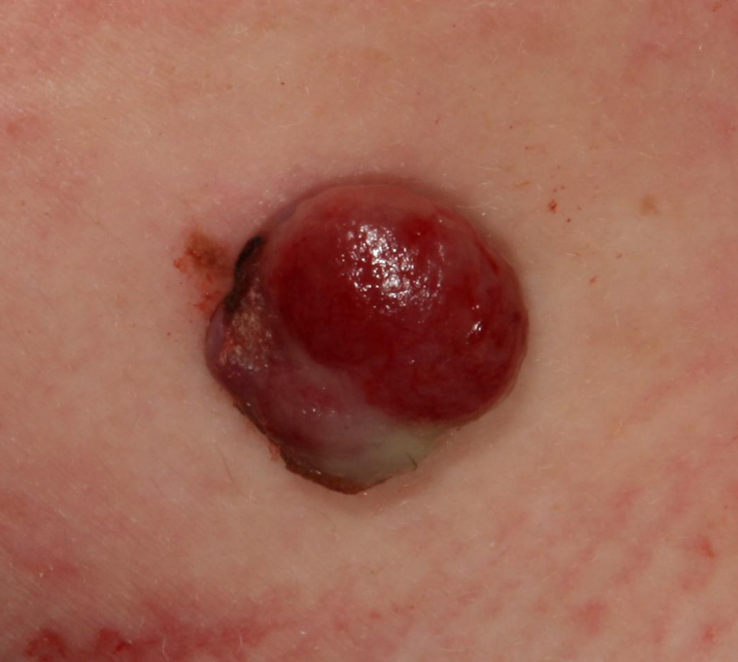
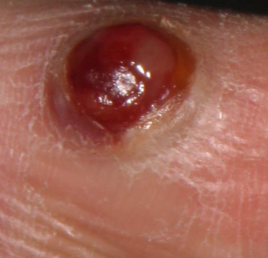
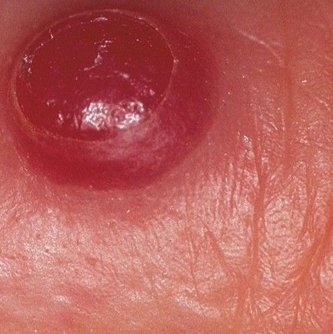
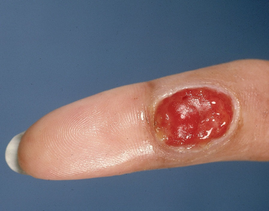
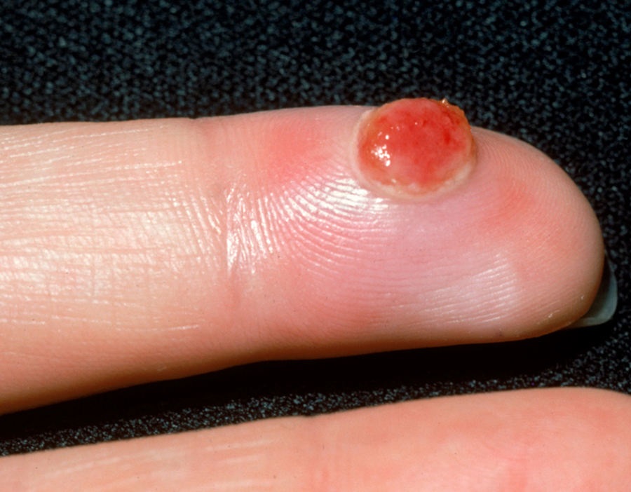
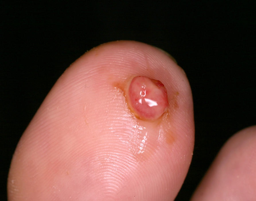
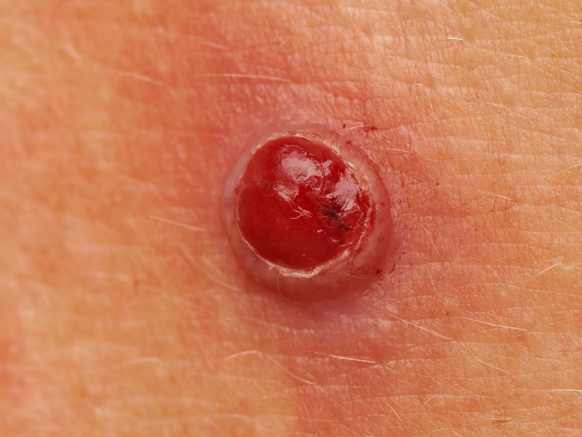
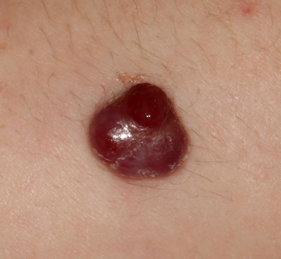
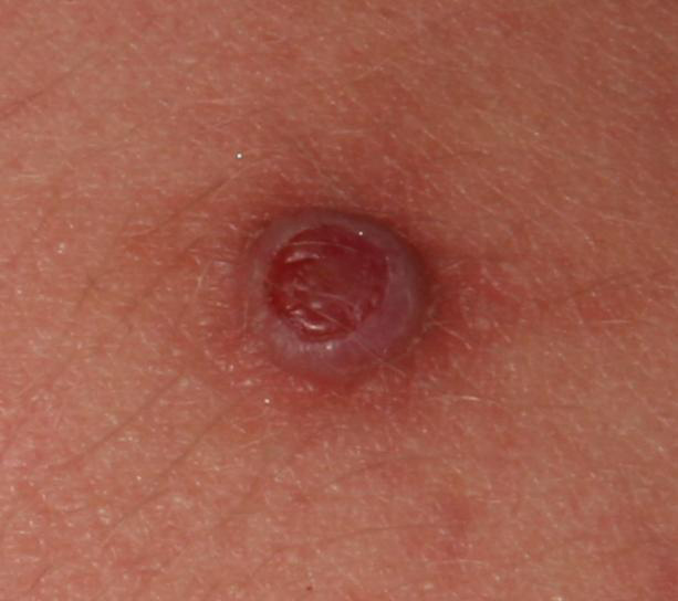
A visual examination of the pyogenic granuloma reveals a hemispherical formation rising above the skin or with a short wide leg, most often symmetrical (oval or round). The surface of the granuloma differs from the texture of ordinary skin: smooth or lobed (such as raspberries), shiny (“wet effect”), with erosion or crusts, with minor injury – it bleeds easily. Large granulomas after infection have become covered with purulent plaque with foci of necrosis.
The boundaries of pyogenic granulomas are clear and even. The edge is represented by the epithelial corolla (the “collar” of the exfoliated epithelium). The color is bright red, cyanotic, cyanotic, in the presence of purulent plaque and foci of necrosis – dirty yellow, gray spots. When pressed, red shades fade.
Hair in the area of pyogenic granuloma usually does not grow.
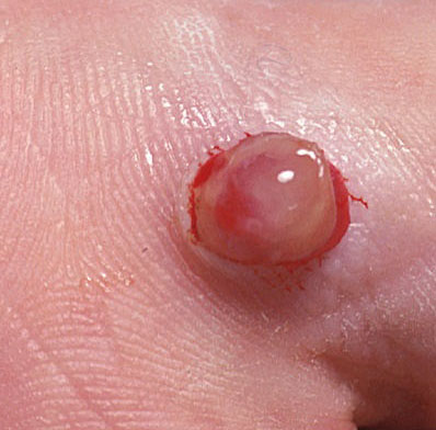
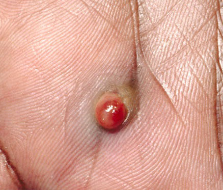
Dimensions are usually 3-15 mm. Formations over 15 mm are rare (with systemic diseases, immunodeficiency states, large burns, pressure sores). Granuloma growth within 1-1.5 cm is usually fast. The height of this above the skin level usually does not exceed 5 mm mm. Spontaneous (spontaneous) regression is rare, more characteristic of pregnant women after delivery.
On palpation of pyogenic granuloma, a soft, elastic, painless nodule is determined. Subjective sensations are also absent. After infection, soreness may appear.
Neoplasms are located mainly of the skin of the hands and feet, especially the palmar and plantar surfaces of the fingers (in the places of the most likely injury and contact with foreign bodies), in the area of the nail ridges (against the background of an ingrown nail), on the face. Other areas, including mucous membranes, are rare (depending on predisposing factors: places of pressure sores, burns, injuries).
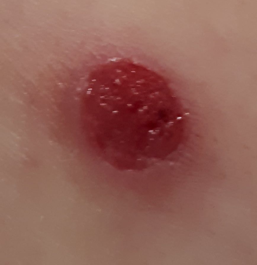
Dermatoscopic Description
With dermatoscopy of papillomatous nevus, the following features are visualized:
- Symptoms of rigidity and filling – a sign of the elasticity of the tumor, which, when compressed, pales and decreases in size;
- After weakening the pressure, the formation acquires the former color and shape;
- A large number of small bright red vascular gaps;
- Lack of vascular pattern (vasculature);
- White whisk;
- Superficial ulceration.
Differential diagnosis
Differential diagnosis is carried out with such formations as:
- Dermatological diseases (pyoderma);
- Spitz Nevus;
- Glomus tumors of the skin;
- Keratoacanthoma;
- Basal cell carcinoma;
- Squamous cell carcinoma;
- Melanoma (especially non-pigmented form);
- Angiosarcoma;
- Kaposi’s Sarcoma;
- Lymphoma of the skin.
Risks
With regard to cancer risks, pyogenic granuloma is safe. This formation does not carry an increased likelihood of malignant tumors. In the absence of external influences (injuries, ultraviolet radiation, ionizing radiation) – the risk of malignancy is comparable to the risk of cancer on unchanged skin.
In the absence of adequate care and treatment, pyogenic granuloma, especially of large sizes, is more dangerous for its complications in the form of infection and suppuration. In these situations, there is a risk of generalization of the infection with corresponding consequences. Also, from large granulomas, if they are damaged, bleeding may develop, which is difficult to cope on their own.
Due to the fact that some malignant neoplasms may be similar in appearance to pyogenic granuloma or appear next to it, there is a certain degree of difficulty in timely differential diagnosis.
Tactics
If pyogenic granuloma is detected, a consultation with a dermatologist or oncologist is recommended. After differential diagnosis and elimination of cancer risks, the possibility of conservative management or the need for treatment is determined. Observation is possible only with small granulomas (a few millimeters), in pregnant women (there is a likelihood of spontaneous involution of education) or in the presence of another, more priority pathology that requires attention. In all other cases, the treatment of pyogenic granuloma is indicated.
In case of refusal of the patient from treatment, active dynamic observation is necessary. At the same time, photo fixation of skin formation is of great value, which will subsequently determine even minor changes in appearance.
Due to the fact that the presence of pyogenic granuloma can make it difficult to timely detect nearby or other tumors that are similar in appearance, but are more dangerous from the oncological point of view, such patients are shown to see a dermatologist or oncologist in the spring and autumn (before the beach season and after him). Of great importance is the mapping of skin neoplasms, which greatly simplifies further observation, the search for new formations, or changes to existing ones.
Treatment
The treatment of pyogenic granuloma is most often surgical: classical excision to the entire thickness of the skin or excision along the plane with the help of an electric or radio scalpel. After removal, a histological examination is mandatory.
When the nature of the formation is not in doubt or a preliminary biopsy is performed with confirmation of the pyogenic granuloma, removal can be performed by laser coagulation, cryo destruction (removal by liquid nitrogen), or electrocoagulation (destruction of the granuloma by an electrocoagulator).
Since pyogenic granuloma is a vascular tumor, active bleeding may occur during removal. In this regard, after removal of the formation, it is necessary to achieve thorough hemostasis.
Prevention
Prevention of the appearance of pyogenic granuloma is a gentle and careful attitude to the skin:
- Exclusion of chronic trauma to the skin and burns;
- Limitation or exclusion of occupational hazards;
- Compliance with safety measures when working with skin-damaging factors;
- Personal hygiene and basic awareness regarding skin neoplasms;
- Timely treatment of infectious diseases, including skin.
It also requires regular examination of the skin, timely consultation with a specialist in the event of external changes in skin tumors, and the removal of potentially dangerous neoplasms.