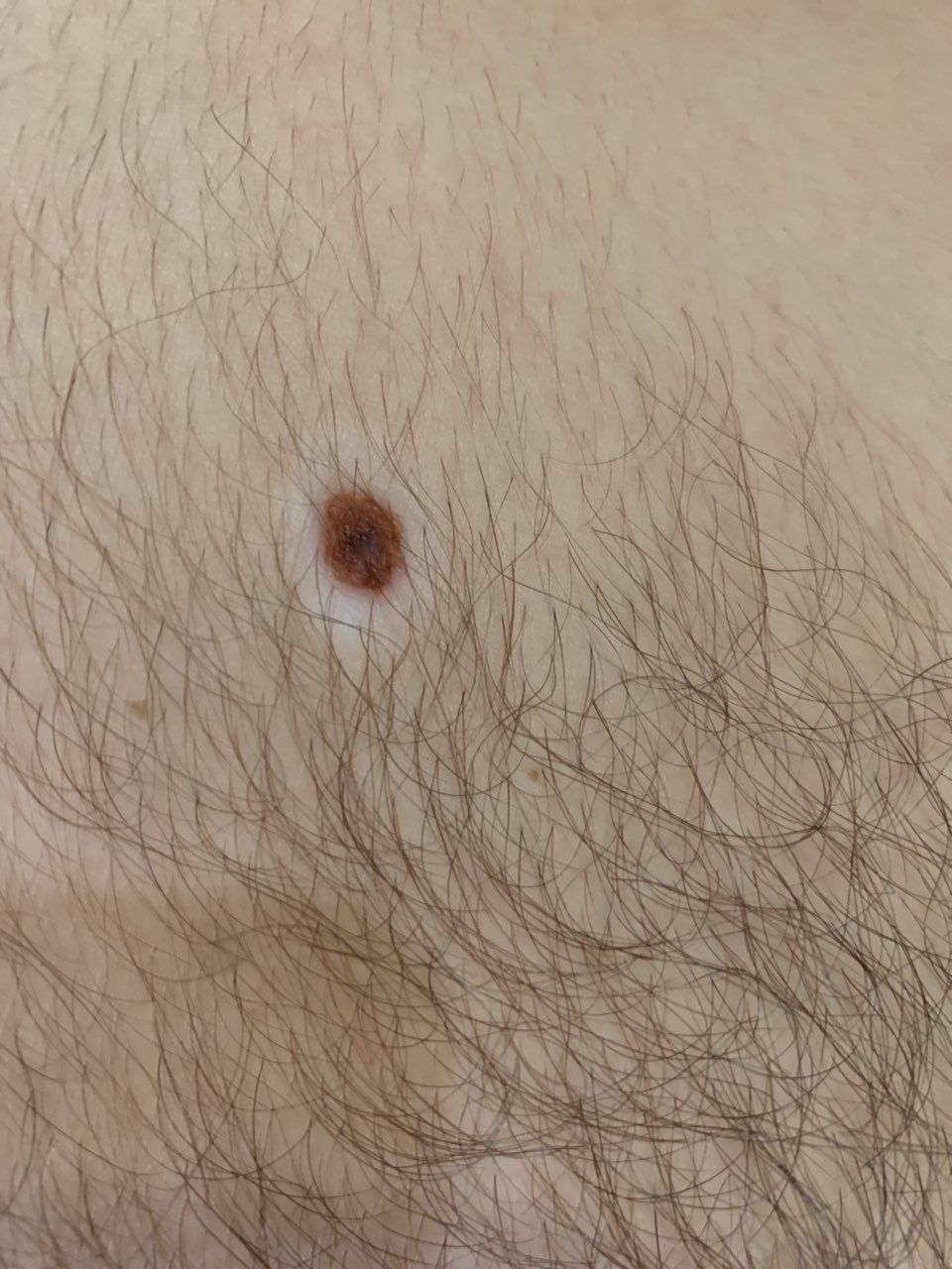
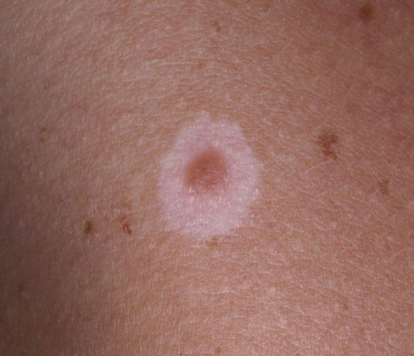
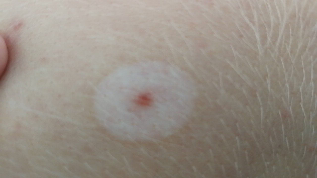
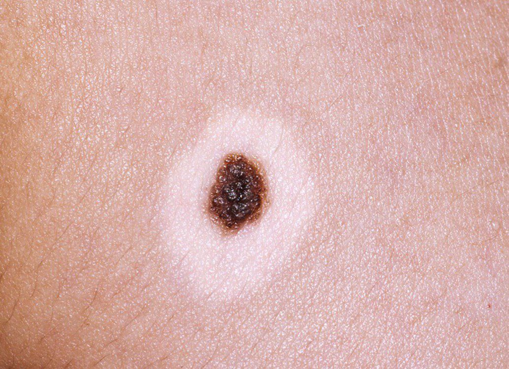
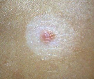
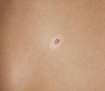
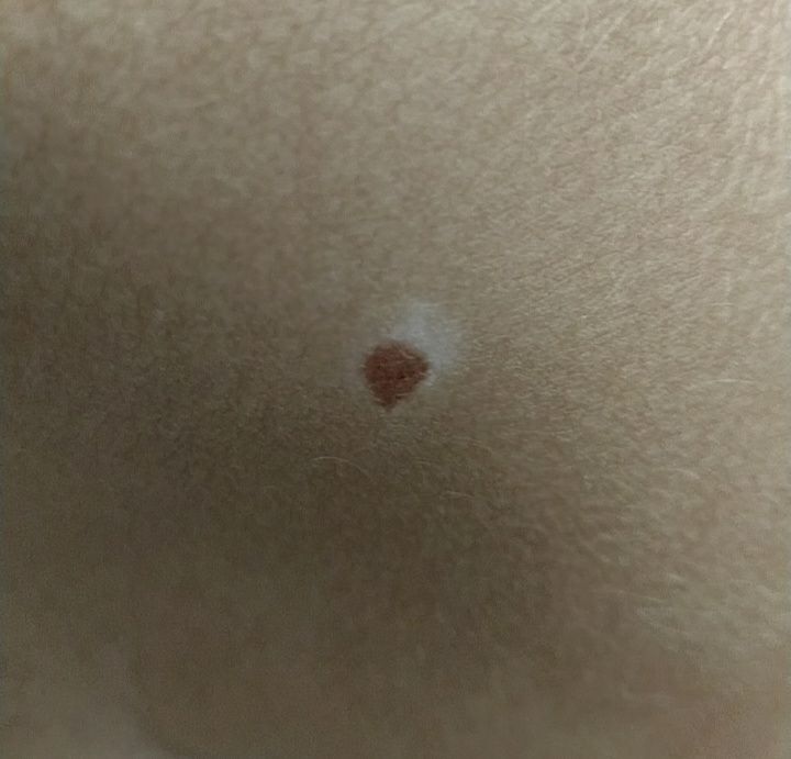
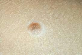
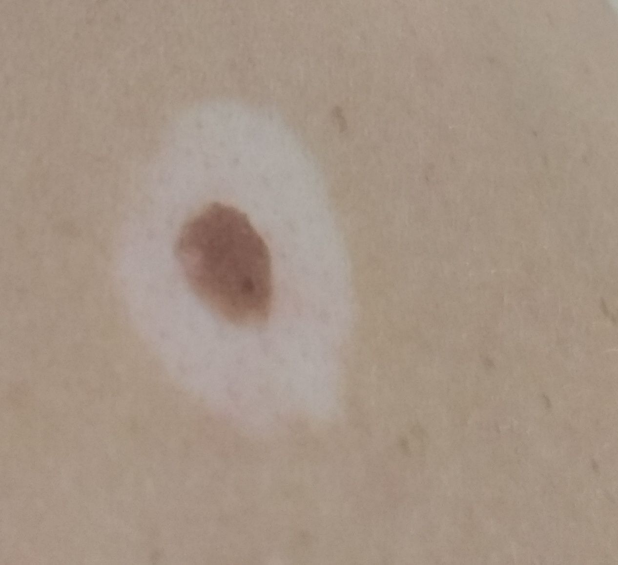
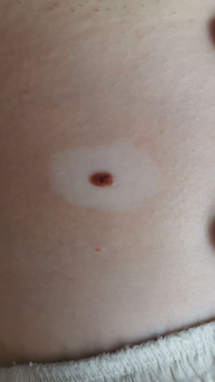
Halo Nevus (Sutton’s Nevus) is a benign neoplasm towering above the skin, surrounded by a rim of hypopigmented skin. Most often, halo nevi are noted in persons 15–25 years of age: first a pigmented, central part appears, around which a colorless ring gradually expands. With age, the reverse involution of the central pigmented part of the nevus can occur (with the preservation of only the focus of hypopigmentation) or with its complete disappearance after 3-4 years.
Predisposing factors
There is no clear reason for the appearance of halo nevus. It is only appropriate to talk about predisposing factors that, to varying degrees, can increase the risk of neoplasms:
- Genetic factor: the appearance of halo nevus may be due to the human genome;
- The presence of vitiligo;
- Ultraviolet radiation: artificial or solar ultraviolet is capable of provoking the appearance of halo nevus;
- Autoimmune diseases: it is submitted that a depigmented ring is due to a secondary localized immune response to regional melanocytes, followed by their destruction.
Diagnostics
Diagnosis of halo nevus is based on a clinical examination, which includes a routine examination of the formation and dermatoscopy. If you suspect malignant growth, a biopsy can be performed.
Symptoms
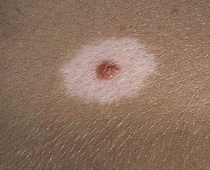
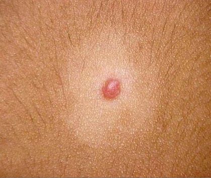
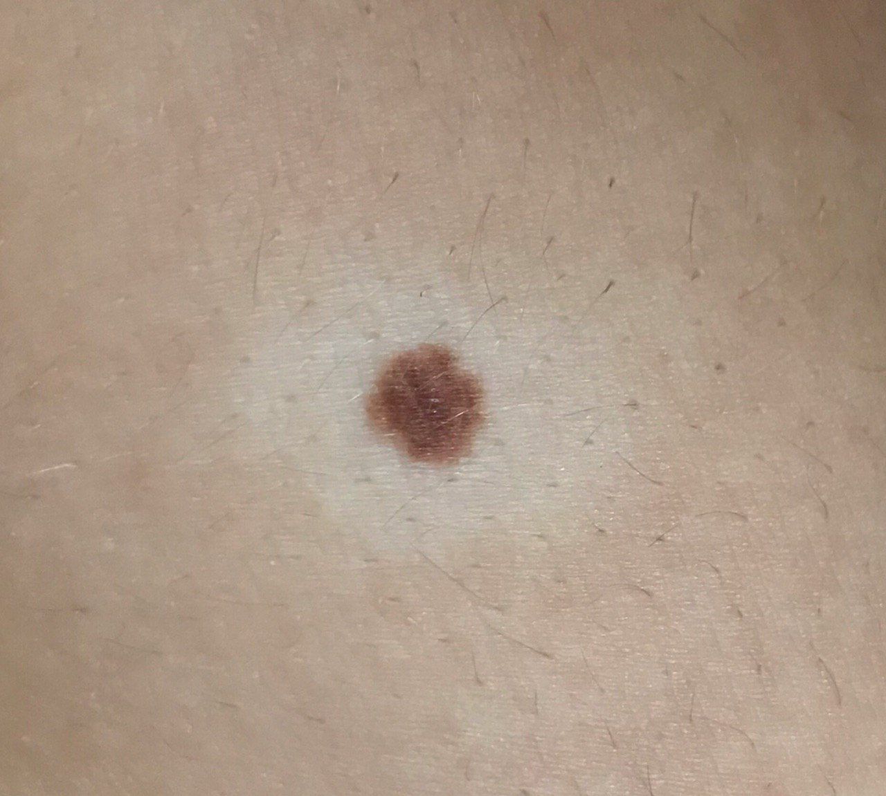
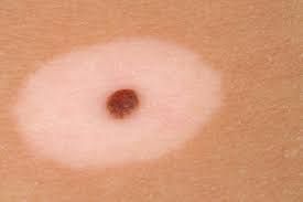
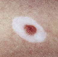
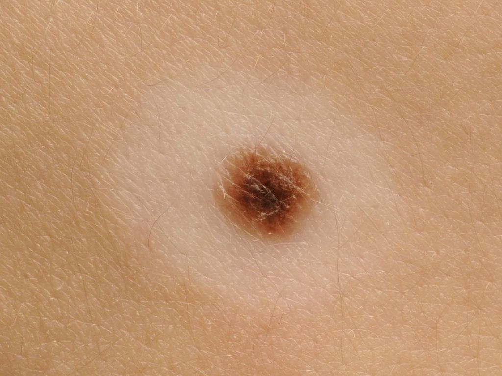
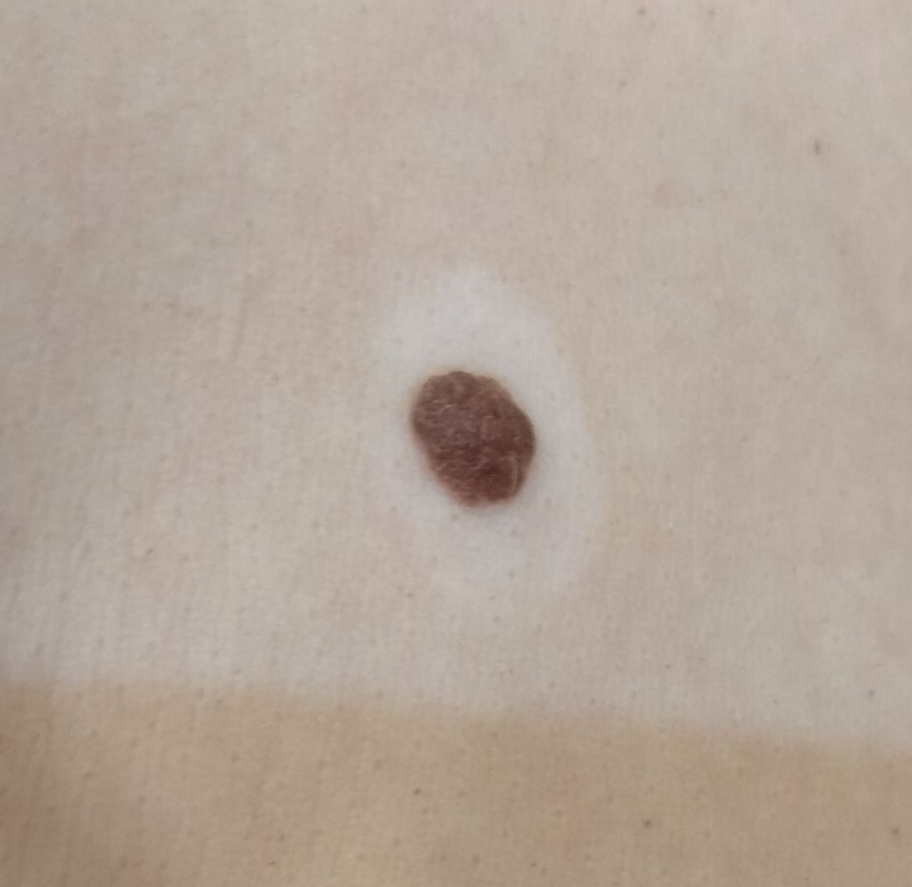
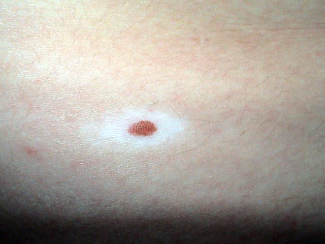
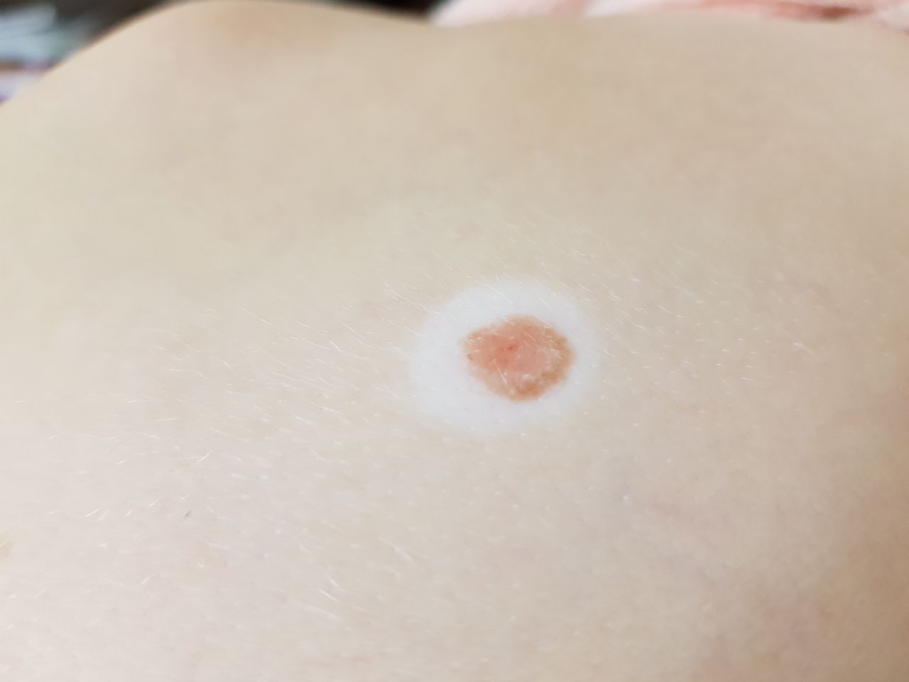
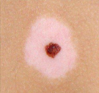
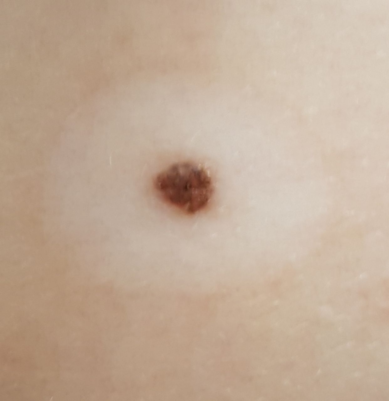
A visual examination of the halo nevus reveals a hemispherical, usually symmetrical (oval or round) formation rising above the skin, around which a rim of depigmented skin is noted. The focus of depigmentation is also regular oval or round in shape, symmetrical.
The surface of the central (pigmented) part of the nevus is slightly different from the texture of the skin or finely tuberous. The surface of the focus of depigmentation is not changed (normal skin pattern).
The borders of the halo nevus are clear and even. The color of the central part varies from flesh, tan to dark brown, while the distribution of pigment throughout the formation is uniform. Sometimes there is a gradual decrease in color intensity in the direction from the center to the periphery, or various shades of the same color within the formation. The color of the surrounding rim is uniform, often completely absent, less often light brown or pale pink or with slight hyperemia, however, the shade is unstable and can change over time. The colorless halo becomes more noticeable and contrast after tanning.
The presence of halo nevus does not affect hair growth. Sometimes in the central part, there is a growth of single coarse bristly or fluffy hair.
The diameter of the central pigmented part of the nevus usually does not exceed 10 mm. The total diameter of the halo nevus, taking into account the depigmented rim, can reach 3-4 cm. Over time, the diameter of the depigmentation focus can vary both in the direction of increase and decrease. The height of the protruding part of the nevus above the skin level usually does not exceed 3-4 mm.
On palpation of the halo nevus, there are no features: the consistency of ordinary skin or slightly softer (central part). Subjective sensations are also absent.
Neoplasms are located mainly on the body, less often – on any part of the body.
Dermatoscopic Description
With dermatoscopy of the central part of the halo nevus, the following features can be visualized:
- Cobblestone street – a network of oval pigment elements;
- Papillary structures – uneven tuberous structures, flattened due to pressure during dermatoscopy;
- Elasticity and deformation under pressure
- Globules – large hyperpigmented ring structures that are evenly distributed throughout the nevus or in the center, are rarely found on the periphery (including gray-brown globules characteristic of hyperkeratosis);
- Spots – hyperpigmented structureless areas located in the center;
- The pigment network is a pattern of hypopigmented holes and homogeneous lines from light brown to dark brown. The lines are evenly thinning to the periphery of the formation;
- Dots – are small hyperpigmented round structures that are located in the center or are found on the pigmented lines of the network;
- Vascular network – represented by slightly curved diffuse monomorphic vessels (regular vasculature);
- Diffuse uniform staining of the entire formation.
With dermatoscopy of the depigmented area – the appearance of ordinary skin with the presence of barely noticeable or no pigment structures at all, perhaps a more marked regular vascular network.
Differential diagnosis
Differential diagnosis is carried out with such neoplasms as:
- Simple nevus
- Spitz Nevus
- Blue nevus
- Vitiligo
- Lichen planus
- Molluscum contagiosum
- Dysplastic nevus
- Basal cell carcinoma
- Melanoma
Risks
Halo nevus is safe and does not carry an increased risk of melanoma. If there is no external effect on such a nevus (trauma, ultraviolet radiation, ionizing radiation), the risk of malignancy is comparable to the risk of a malignant tumor on unchanged skin. Signs of a possible malignancy: a change in appearance, the appearance of subjective sensations.
Tactics
In the absence of any damaging effect on the halo nevus, changes in appearance and subjective sensations, self-control (or inspection with the help of other persons in inaccessible areas) is sufficient at least once a year. If mechanical damage to the nevus has occurred, its active irradiation with ultraviolet or ionizing radiation, as well as if any changes in the nevus itself are noticed or previously absent sensations appear, you need to consult a dermatologist or oncologist.
The specialist determines the possibility of further dynamic monitoring (terms are determined individually) or indications are made for removal of the damaged nevus. It is necessary to remove those nevi that are subject to constant, chronic trauma to clothing, jewelry, or due to the characteristics of professional employment.
In the case of dynamic observation, photo fixation of skin neoplasms is of great value, which will subsequently determine even minor changes in the appearance of the nevus.
Patients with multiple moles are shown an examination by a dermatologist or oncologist in the spring and autumn (before and after the beach season). Such patients are also recommended to compile a map of skin neoplasms, which greatly simplifies further observation, the search for new formations, or a change in existing ones.
Treatment
Only surgical (classic, using an electric or radio scalpel) with a mandatory histological examination.
Treatment of halo nevus using destructive methods (laser removal or cryodestruction) is not recommended.
Prevention
Prevention of the appearance of nevi and their malignancy consists in a gentle and careful attitude to the skin:
- Limitation of ultraviolet radiation (tanning bed, solar tanning);
- The use of protective creams during periods of active sun;
- Exclusion of chronic skin trauma
- Limitation or exclusion of ionizing radiation, occupational hazards;
- Compliance with safety measures when working with skin-damaging factors;
- Personal hygiene and basic awareness of skin tumors.
Also, regular inspection of halo nevus, timely consultation of a specialist in the event of external changes, and the removal of potentially dangerous neoplasms are necessary.