Spitz Nevus (Epithelioid and Spindle-Cell Nevus) – a benign neoplasm towering above the skin. As a rule, Spitz nevus is acquired (in 10% – congenital). About 70% of all cases are observed in people under the age of 20 years. Multiple lesions are noted sometimes. By gender, nevi are equally common among male and female patients.
Predisposing factors
There is no clear reason for the appearance of Spitz nevi. It is only appropriate to talk about predisposing factors that, to varying degrees, can increase the risk of neoplasms:
- Genetic factor: the appearance of Spitz nevus may be due to the human genome;
- Ultraviolet radiation: artificial or solar ultraviolet can provoke the appearance of new nevi;
- Hormonal changes: especially during puberty or during pregnancy.
Diagnostics
Diagnosis of Spitz nevus is based on a clinical examination, which includes a routine examination of the formation and dermatoscopy. If you suspect malignant growth, a biopsy can be performed.
Symptoms
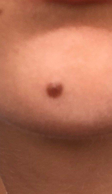
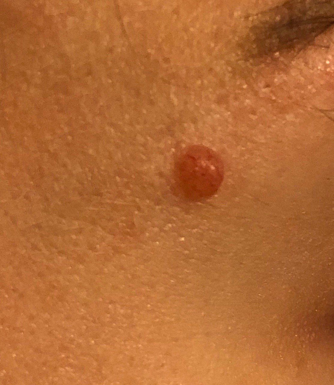
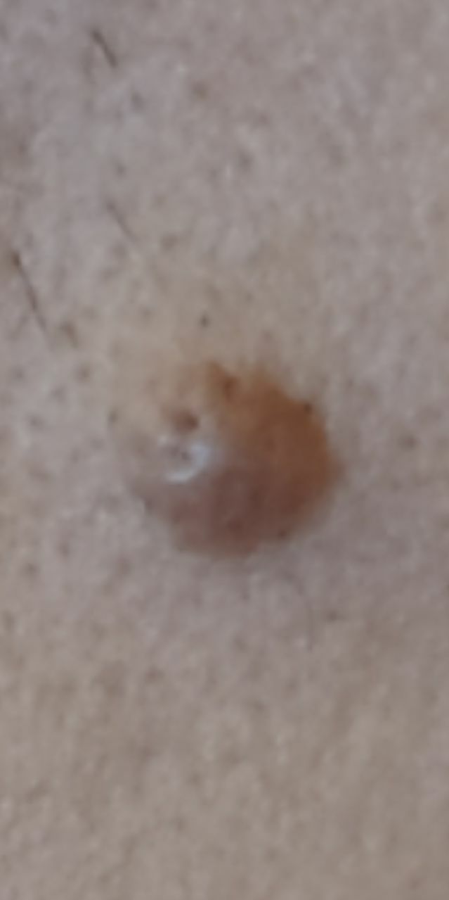
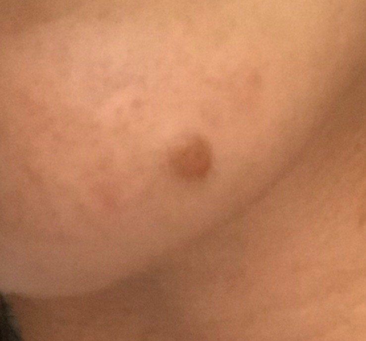
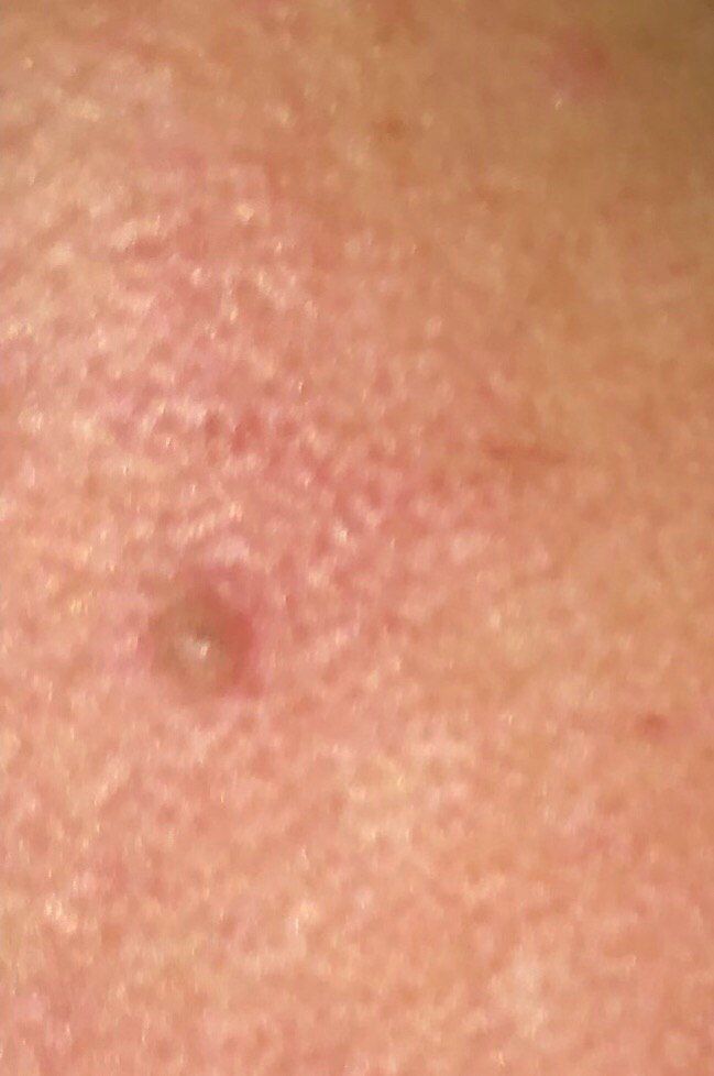
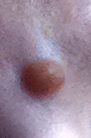
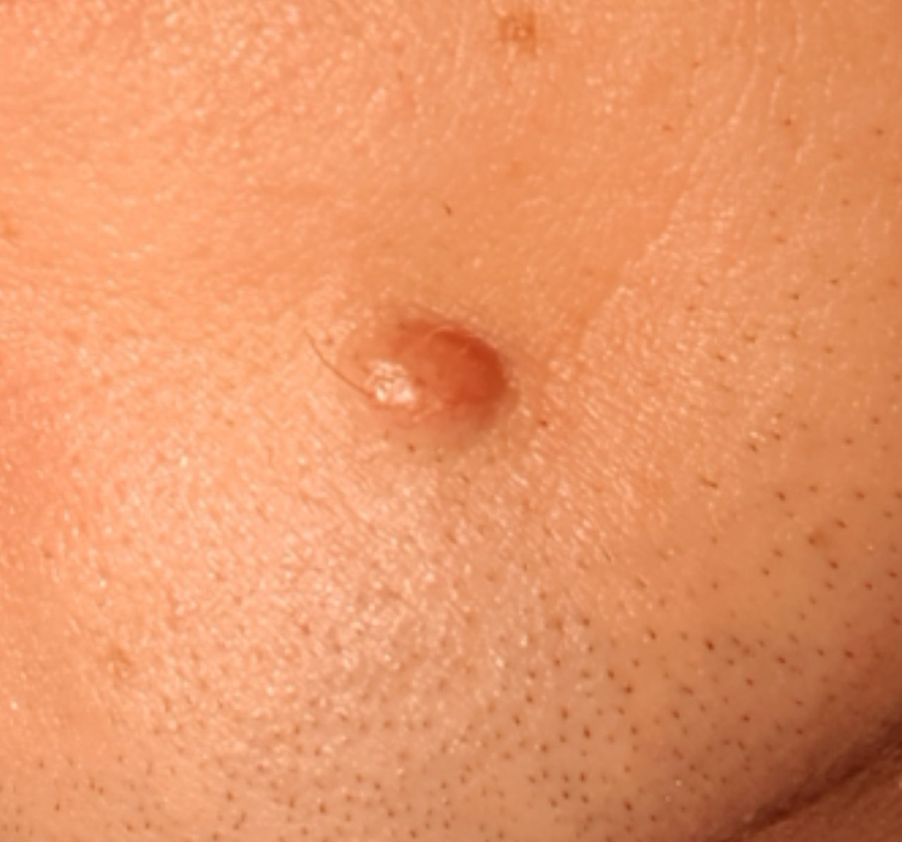
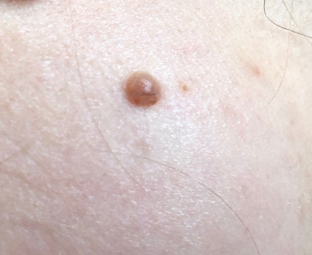
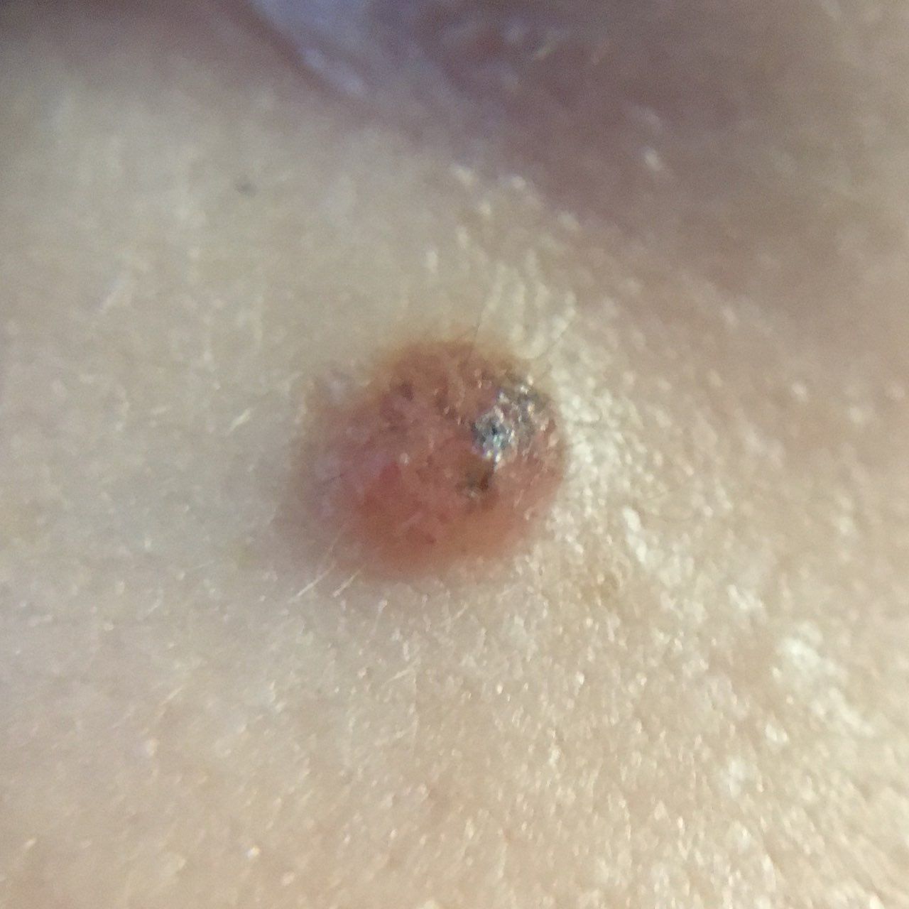
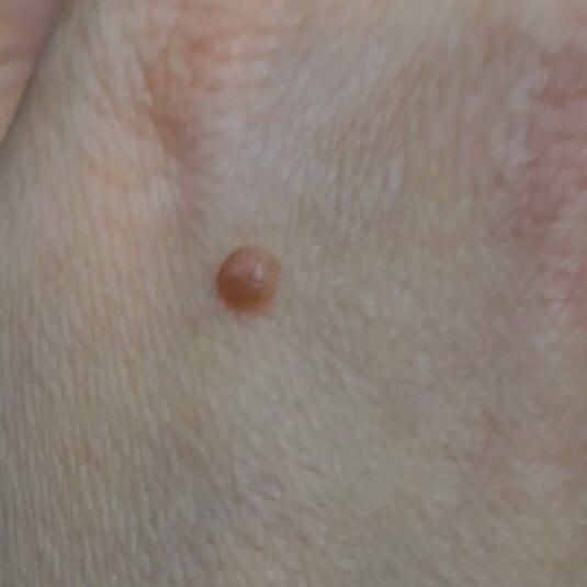
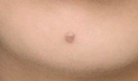
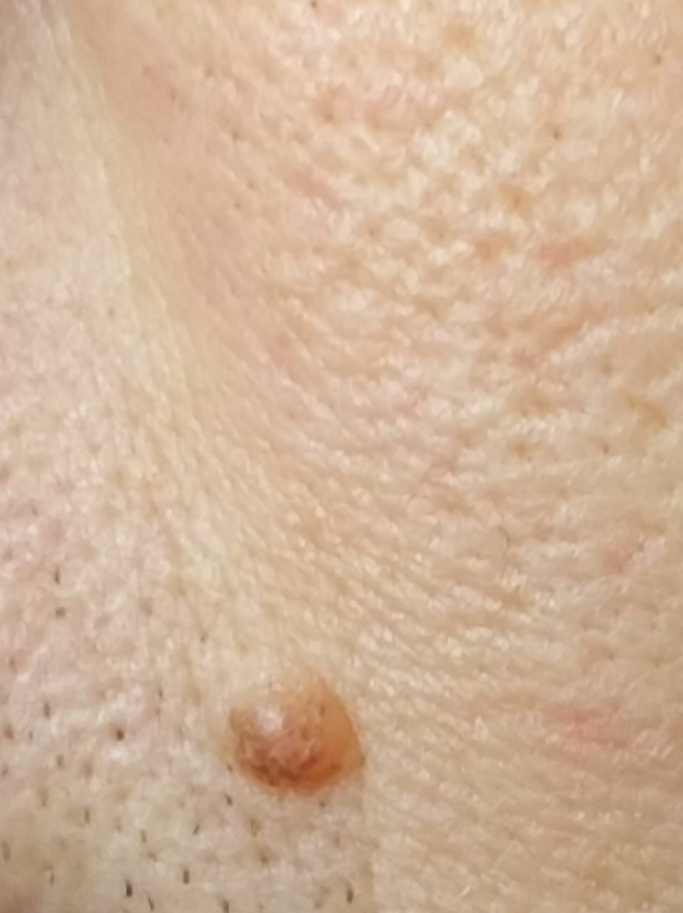
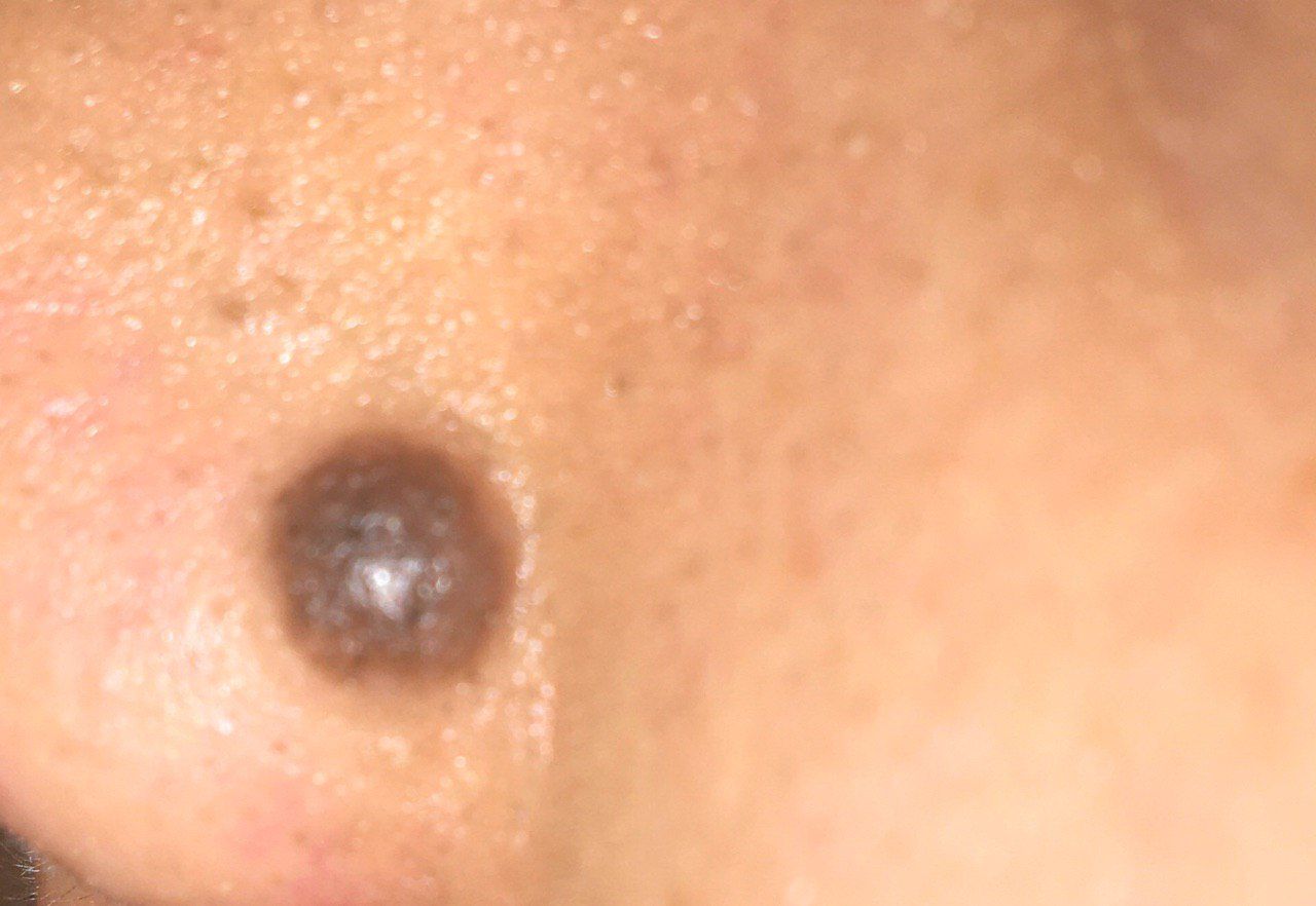
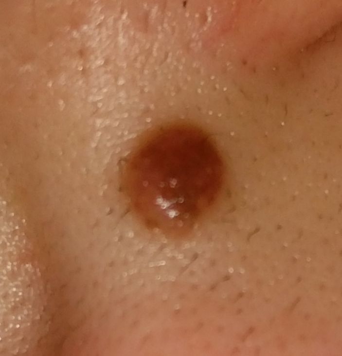
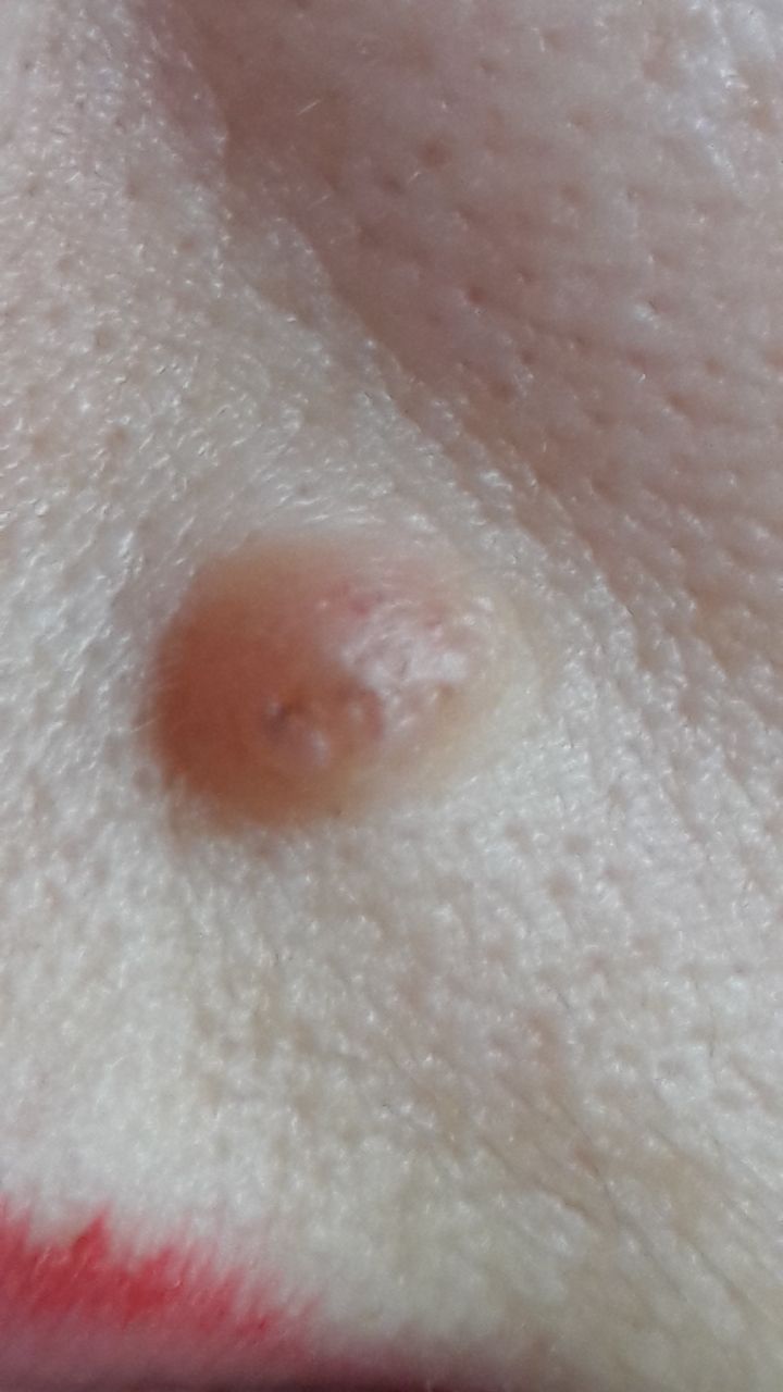
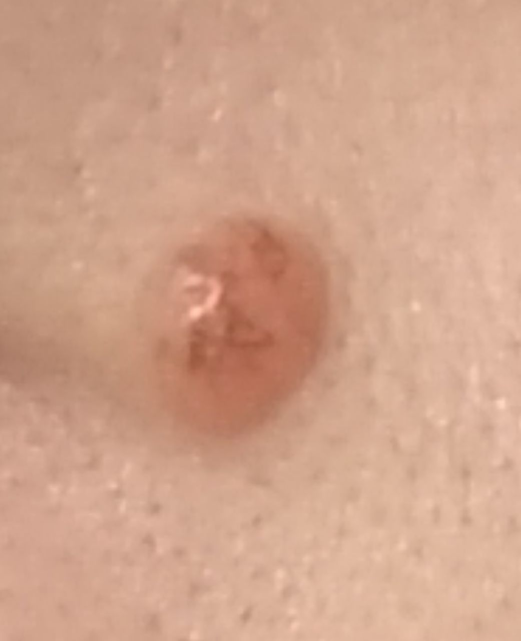
A visual examination of the Spitz nevus determines a hemispherical or flattened (rarely flat) formation rising above the skin, most often symmetrical (oval or round). The surface of the nevus is slightly different from the texture of ordinary skin or finely tuberous.
The borders of the Spitz Nevus are clear and even. The color of a simple nevus varies from intense-bodily (light red) to brown or dark brown, while the distribution of pigment throughout the formation is uniform. Sometimes there is a gradual decrease in color intensity in the direction from the center to the periphery, or various shades of the same color within the nevus.
In the area of Spitz nevus, hair never grows, which is a distinguishing feature of this formation and plays a significant role in differential diagnosis.
The diameter of the Spitz nevus is usually 3-8 mm. The height above the skin usually does not exceed 5-7 mm.
On palpation, the nevus is slightly denser than normal skin. There are no subjective sensations.
They are located mainly on the face, neck, and limbs, less often – on the body.
Dermatoscopic Description
With dermatoscopy, the following Spitz nevus features are visualized:
- Star pattern: the presence of pigment strips, dots and / or globules radially diverging from the center to the periphery – the main dermatoscopic symptom;
- Symmetric blue-white structure against the background of pigmented elements (often spots) in the center of the nevus;
- Elasticity and deformation under pressure;
- Vascular network – represented by slightly curved diffuse monomorphic vessels (regular vasculature);
- Diffuse uniform staining of the entire formation.
Differential diagnosis
Differential diagnosis is carried out with such pigmented neoplasms, as:
- Simple nevus
- Papillomatous nevus
- Molluscum contagiosum
- Blue nevus
- Dysplastic nevus
- Basal cell carcinoma
- Melanoma
Risks
Spitz Nevus is safe and does not carry an increased risk of melanoma. In the absence of external effects on such a nevus (trauma, ultraviolet radiation, ionizing radiation), the risk of malignant degeneration is comparable to the risk of melanoma on unchanged skin.
Signs of a possible malignancy: a change in appearance, the appearance of subjective sensations. The risk of melanoma on the background of congenital nevus is less than 1%.
Tactics
In the absence of any damaging effect on the Spitz nevus, changes in appearance and subjective sensations – self-control (or examination with the help of other persons in inaccessible areas) is enough at least once a year. If mechanical damage to the nevus has occurred, its active irradiation with ultraviolet or ionizing radiation, as well as if any changes in the nevus itself are noticed or previously absent sensations appear, you need to consult a dermatologist or oncologist.
The specialist determines the possibility of further dynamic monitoring (terms are determined individually) or indications are made for removal of the damaged nevus. It is necessary to remove those nevi that are subject to constant, chronic trauma to clothing, jewelry, or due to the characteristics of professional employment.
In the case of dynamic observation, photo fixation of skin neoplasms is of great value, which will subsequently determine even minor changes in the appearance of the nevus.
Patients with multiple moles require an examination by a dermatologist or oncologist in the spring and autumn (before and after the beach season). Such patients are also recommended to compile a map of skin neoplasms, which greatly simplifies further observation, the search for new formations, or a change in existing ones.
Treatment
Only surgical (classic, using an electric or radio scalpel) with a mandatory histological examination.
Treatment of Spitz nevus using destructive methods (laser removal or cryodestruction) is not recommended.
Prevention
Prevention of the appearance of nevi and their malignancy consists in a gentle and careful attitude to the skin:
- Limitation of ultraviolet radiation (tanning bed, solar tanning);
- The use of protective creams during periods of active sun;
- Exclusion of chronic skin trauma;
- Limitation or exclusion of ionizing radiation, occupational hazards;
- Compliance with safety measures when working with skin-damaging factors;
- Personal hygiene and basic awareness of skin tumors.
Also, a regular examination of Spitz nevi is necessary, timely consultation of a specialist in the event of external changes and removal of potentially dangerous neoplasms.