Papillomatous nevus (benign nevus, pigmented nevus, mole) is a benign neoplasm towering above the skin. As a rule, papillomatous nevus is acquired. With age, the number of formations increases, the peak of occurrence is noted at 15-30 years. This type of nevus is characterized by multiplicity, the proportion of which increases with age. By gender, nevi are somewhat more common in women compared with men, in a ratio of 3: 2, respectively.
Predisposing factors
There is no clear reason for the appearance of papillomatous nevi. It is only appropriate to talk about predisposing factors that, to varying degrees, can increase the risk of neoplasms:
- Genetic factor: the appearance of papillomatous nevus may be due to the human genome;
- Ultraviolet radiation: artificial or solar ultraviolet leads to faster reproduction of nevoid cells (nevus cells) and excessive production of melanin (pigment, the accumulation of which is noted in nevus);
- Ultraviolet radiation: artificial or solar ultraviolet leads to faster reproduction of nevoid cells (nevus cells) and excessive production of melanin (pigment, the accumulation of which is noted in nevus);
- Ionizing radiation, viral diseases, and injuries can also provoke the appearance or growth of papillomatous nevi.
Diagnostics
Diagnosis of papillomatous nevi is based on a clinical examination, which includes a routine examination of the formation and dermatoscopy. If you suspect malignant growth, a biopsy can be performed.
Symptoms
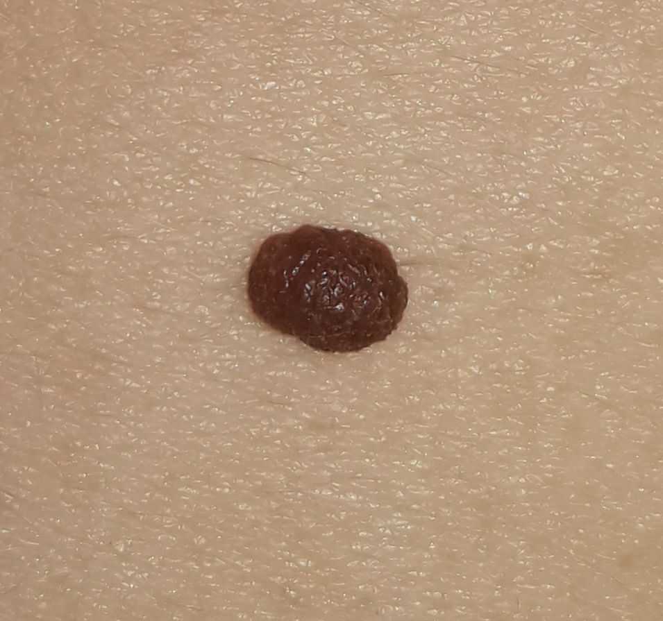
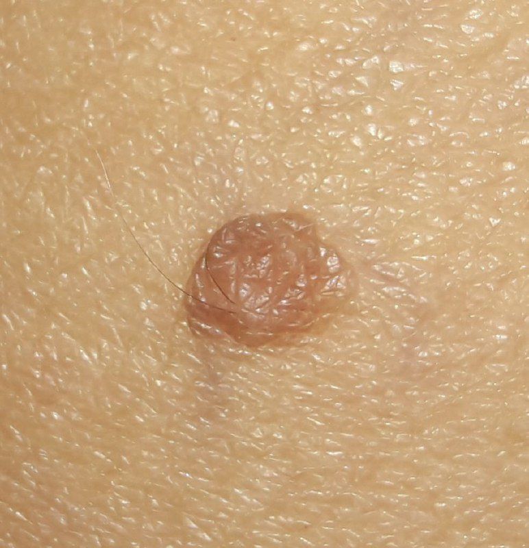
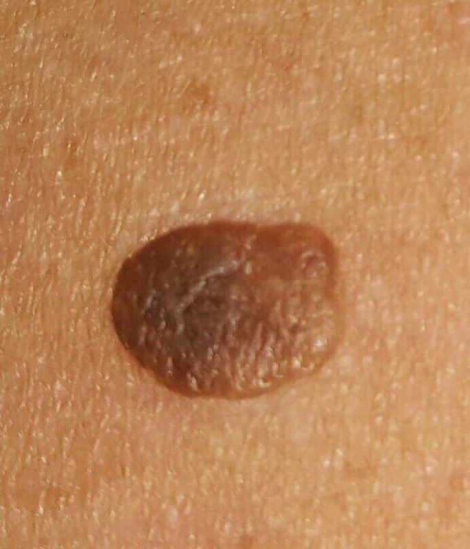
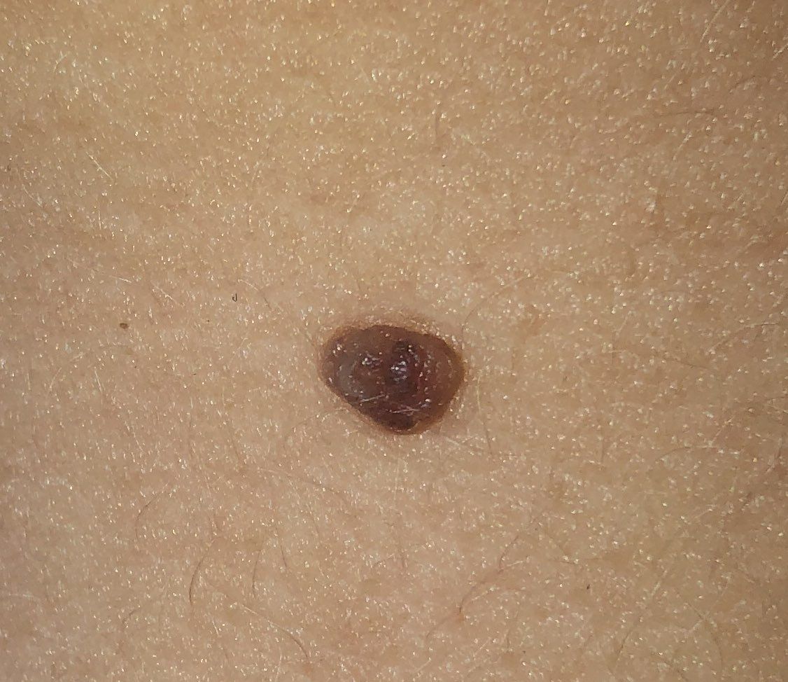
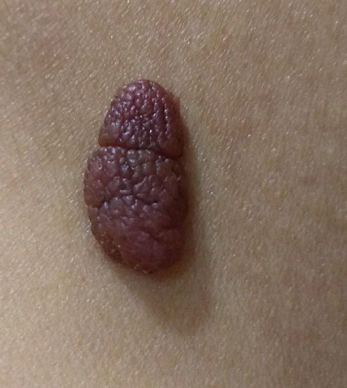
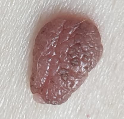
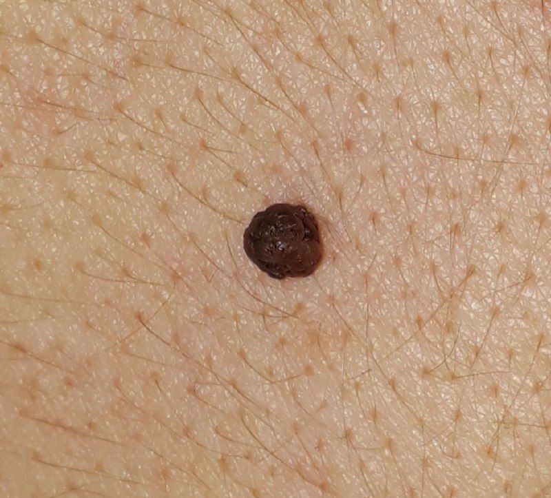
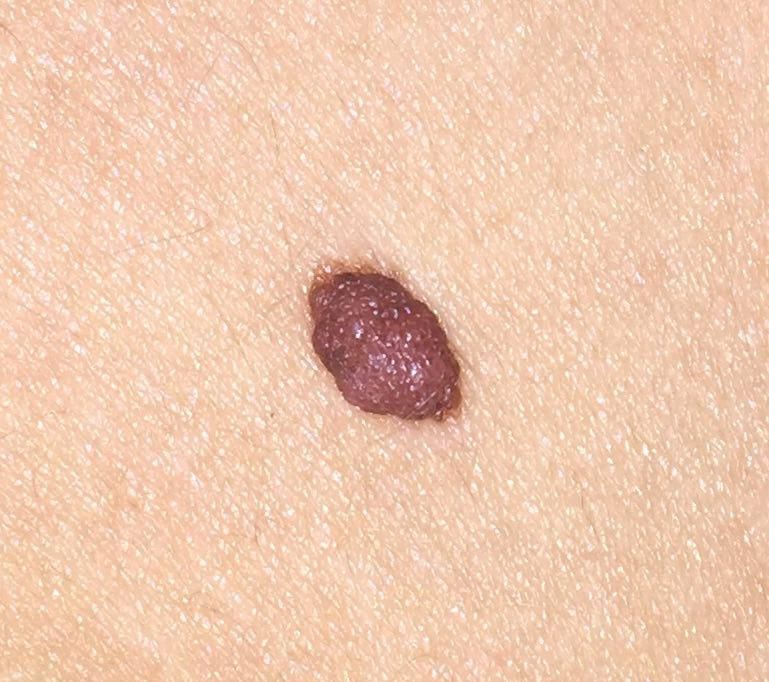
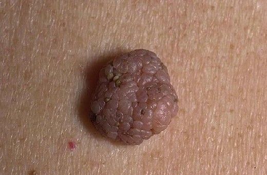
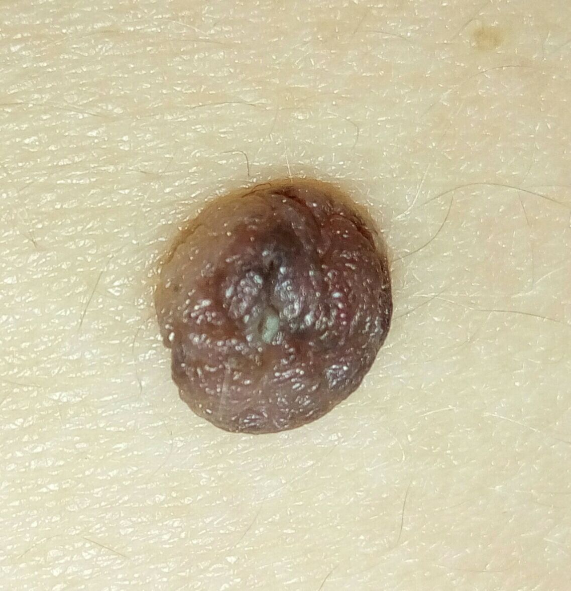
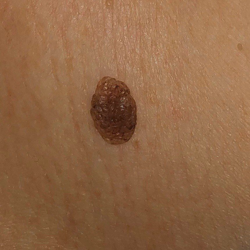
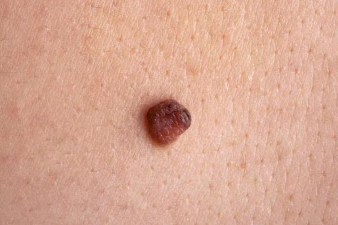
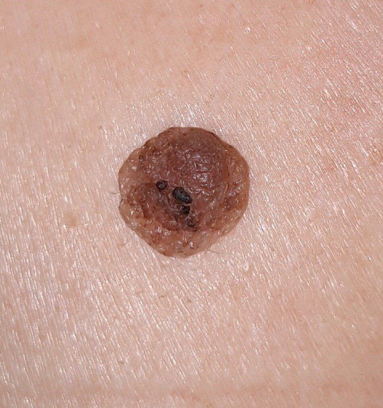
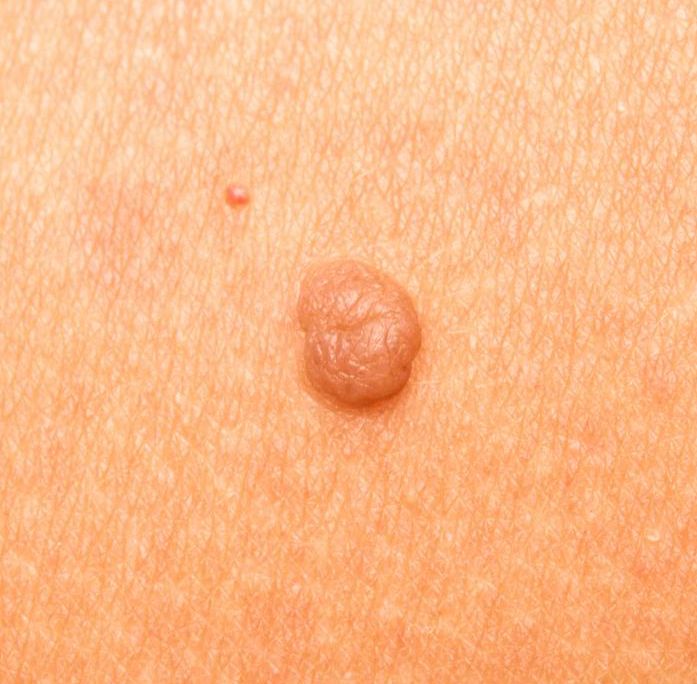
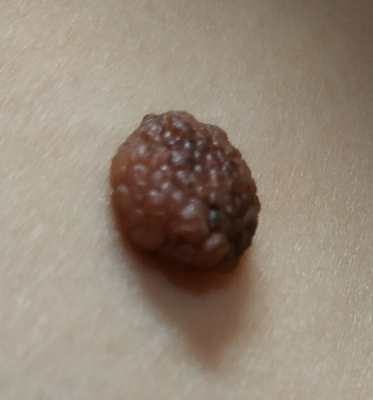
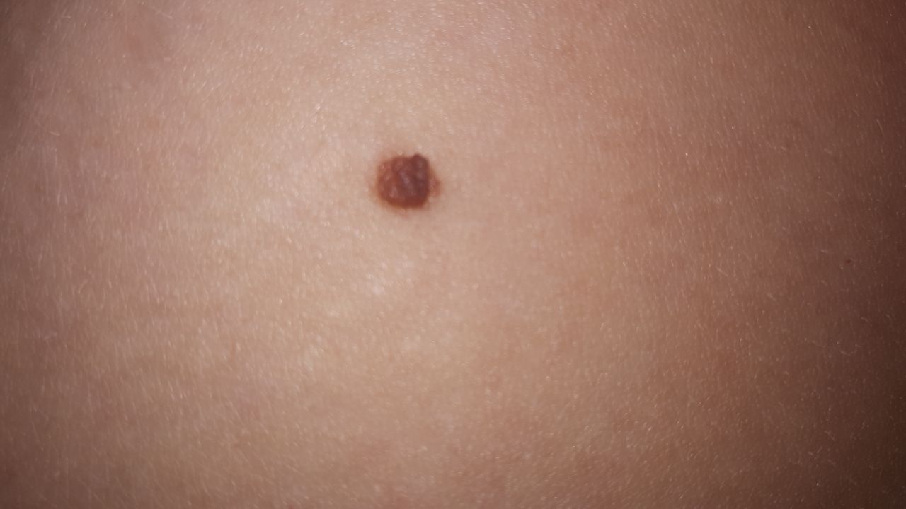
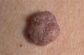
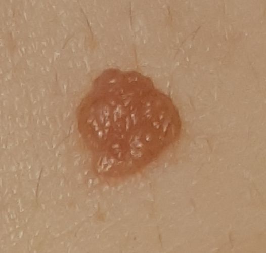
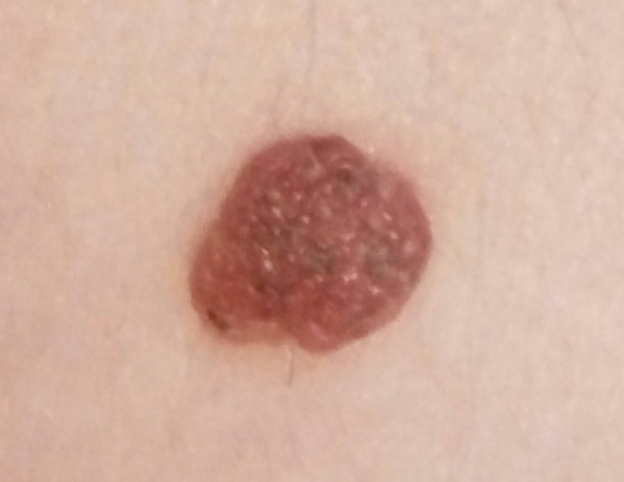
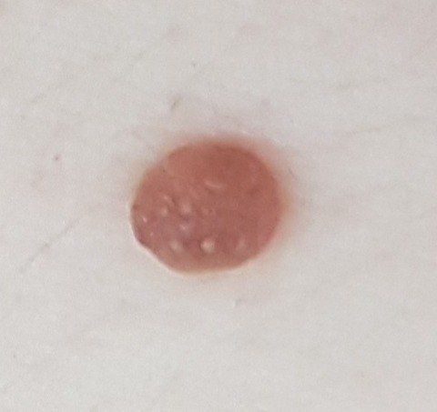
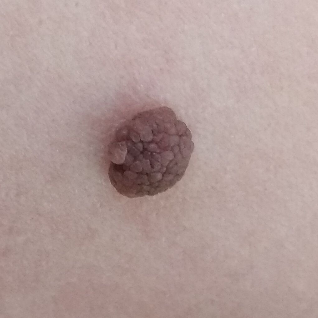
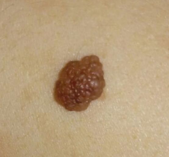
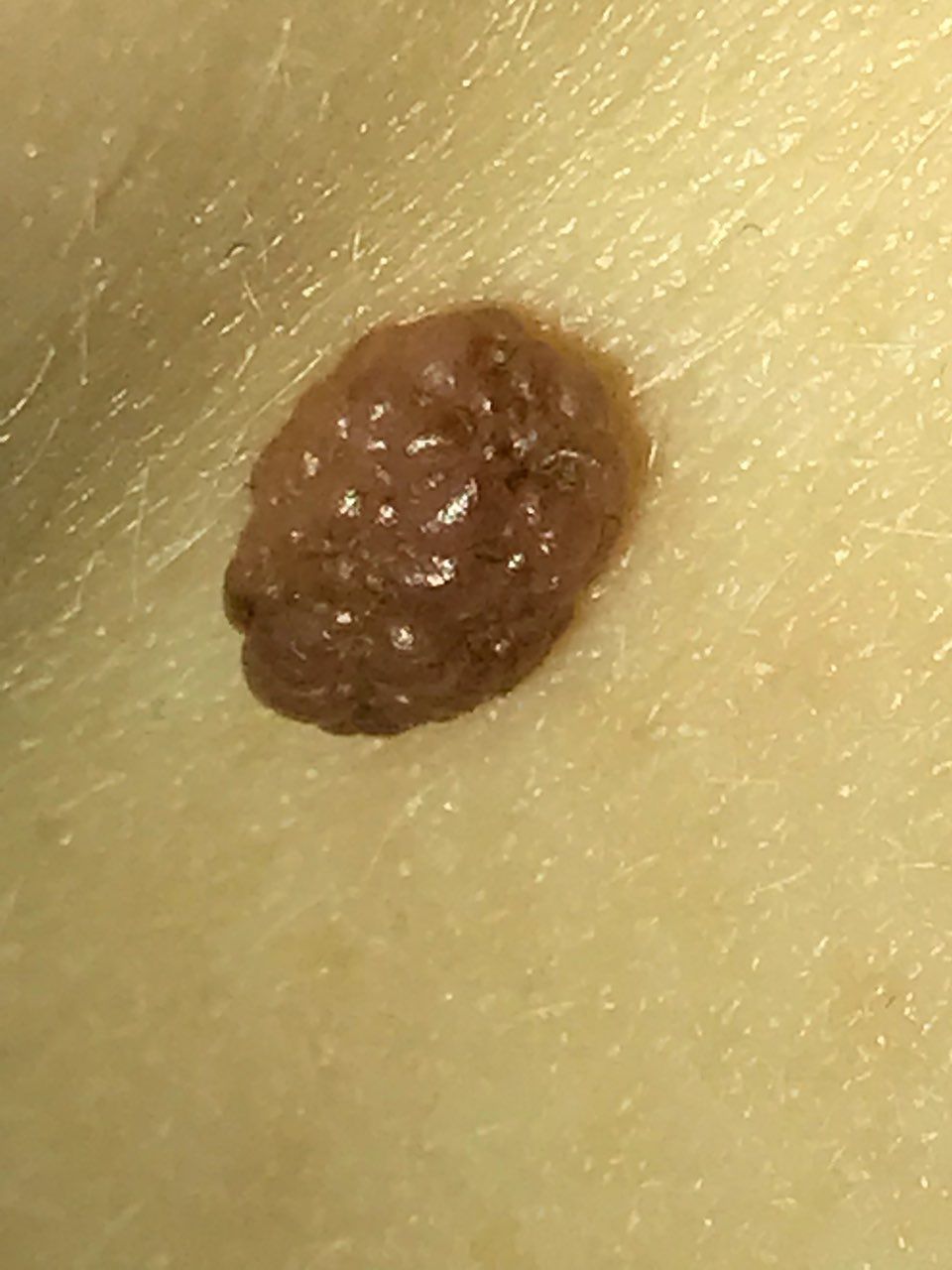
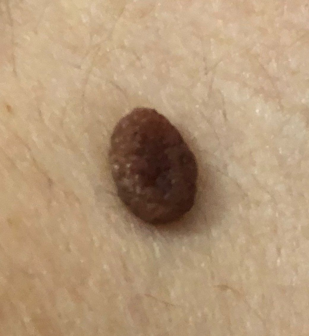
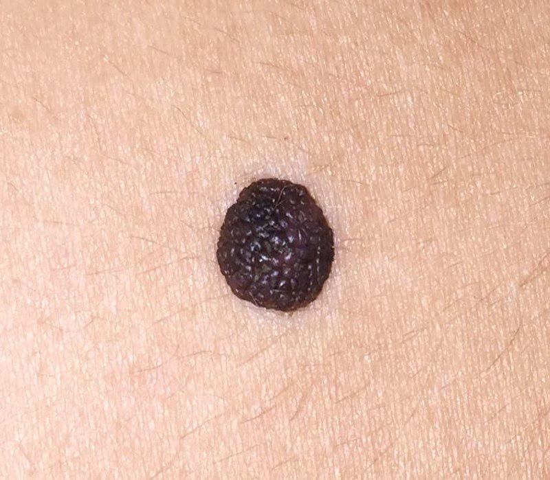
A visual examination of the papillomatous nevus reveals a hemispherical formation rising above the skin or with a short wide leg, most often symmetrical (oval or round). Large nevi can be irregular in shape. The surface of the nevus is slightly different from the texture of ordinary skin or finely tuberous. Sometimes the papillomatous nevus looks like a coarse warty formation (the surface in the form of large, uneven papillae is the verrucous nevus), which is more typical for nevi from 8 mm or more.
The borders of the papillomatous nevus are clear and even. Large nevi can be with uneven edges. The color of a simple nevus varies from flesh, tan to dark brown (almost black) in color, while the distribution of pigment throughout the formation is uniform. Sometimes there is a gradual decrease in color intensity in the direction from the center to the periphery or different shades of the same color within the same formation (typical for verrucous nevus).
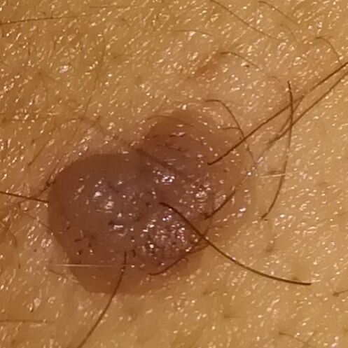
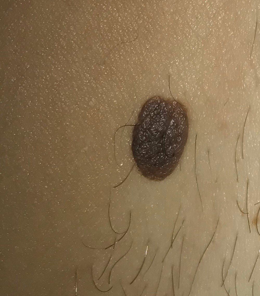
The presence of benign nevus does not affect hair growth. Less often in the area of congenital nevus, there is a more intensive growth of coarse bristly hair, which is usually combined with pronounced brown pigmentation, or vice versa, downy hair (combined with hypopigmented papillomatous nevi).
The size of the nevi can vary within wide limits, however, the most common formations are up to 15 mm. Papillomatous nevi over 15 mm are rare. The height of such a nevus above the skin level usually does not exceed 10 mm. Papillomatous nevi in the form of cauliflower and much larger sizes are rare.
On palpation of a simple nevus, there are no features: the consistency of ordinary skin (larger formations) or slightly softer (formations up to 5 mm). Subjective sensations are also absent.
Neoplasms are located mainly on the face, scalp, neck, trunk, and less commonly on the limbs.
Dermatoscopic Description
With dermatoscopy of a papillomatous nevus, the following features are visualized:
- Cobblestone street – a network of oval pigment elements;
- Papillary structures – uneven tuberous structures, flattened due to pressure during dermatoscopy;
- Elasticity and deformation under pressure (as well as the first two points) are characteristic dermatoscopic signs of papillomatous nevi;
- Globules – large hyperpigmented ring structures that are evenly distributed throughout the nevus or in the center, are rarely found on the periphery (including gray-brown globules characteristic of hyperkeratosis);
- Spots – hyperpigmented structureless areas located in the center;
- Vascular network – represented by slightly curved diffuse monomorphic vessels (regular vasculature);
- Diffuse uniform staining of the entire formation.
Differential diagnosis
Differential diagnosis is carried out with pigmented neoplasms such as:
- Nevus of the sebaceous glands
- Halo nevus
- Spitz Nevus
- Blue nevus
- Dysplastic nevus
- Melanoma
Risks
Papillomatous nevus is safe and does not carry an increased risk of melanoma. In the absence of external effects on such a nevus (trauma, ultraviolet radiation, ionizing radiation), the risk of malignant degeneration is comparable to the risk of melanoma on unchanged skin. Signs of a possible malignancy: a change in appearance, the appearance of subjective sensations.
A slightly increased risk of melanoma is observed in congenital nevus, but this is more typical for large pigmented lesions (more than 20 cm in diameter). The risk of melanoma on the background of congenital nevus up to 20 cm is less than 1%.
Tactics
In the absence of any damaging effect on the papillomatous nevus, changes in appearance and subjective sensations, self-control (or examination with the help of other persons in inaccessible areas) is enough at least once a year. If mechanical damage to the nevus has occurred, its active irradiation with ultraviolet or ionizing radiation, as well as if any changes in the nevus itself are noticed or previously absent sensations appear, you need to consult a dermatologist or oncologist.
The specialist determines the possibility of further dynamic monitoring (terms are determined individually) or indications are made for removal of the damaged nevus. It is necessary to remove those nevi that are subject to constant, chronic trauma to clothing, jewelry, or due to the characteristics of professional employment.
In the case of dynamic observation, photo fixation of skin neoplasms is of great value, which will subsequently determine even minor changes in the appearance of the nevus.
Patients with multiple moles require an examination by a dermatologist or oncologist in the spring and autumn (before and after the beach season). Such patients are also recommended to compile a map of skin neoplasms, which greatly simplifies further observation, the search for new formations, or a change in existing ones.
Treatment
Only surgical (classic, using an electric or radio scalpel) with a mandatory histological examination.
Treatment of pigmented papillomatous nevus using destructive methods (laser removal or cryodestruction) is not recommended.
Prevention
Prevention of the appearance of nevi and their malignancy consists in a gentle and careful attitude to the skin:
- Limitation of ultraviolet radiation (tanning bed, solar tanning);
- The use of protective creams during periods of active sun
- Exclusion of chronic skin trauma;
- Limitation or exclusion of ionizing radiation, occupational hazards;
- Compliance with safety measures when working with skin-damaging factors;
- Personal hygiene and basic awareness of skin tumors.
Also, regular inspection of pigmented papillomatous nevi, timely consultation of a specialist in the event of external changes, and the removal of potentially dangerous neoplasms are necessary.